
Visualizing Female Meiosis in Arabidopsis
In plants, compared to male meiosis, female meiosis has remained largely unexplored. In our recent study published in The Plant Cell (Hu and Prusicki et al., 2026), by live cell imaging, we visualize female meiosis in the model plant Arabidopsis thaliana, advancing the knowledge of reproductive development.
Background: To sexually reproduce, multicellular organisms have to reduce their chromosome number by half so that the genomic content does not increase over generations. This is accomplished by a special cell division called meiosis. While much has been learned about male meiosis, which, in the case of the flowering plant Arabidopsis, takes place in anthers, little is known about female meiosis, which is executed in ovule primordia. The reason for this is that the cells undergoing meiosis in the female flower parts are deeply embedded in maternal tissues and hence very poorly accessible.
Question: If at all, female meiosis has up to now been solely studied by static methods such as the staining of chromosomes with dyes and/or the detection of proteins with antibodies. While these techniques were and are still important, they hardly deliver information about the dynamics of meiosis, such as the chromosome movements and changes in abundance of proteins over time.
Findings: Here, we have used the model plant Arabidopsis thaliana and established a live-cell imaging system to follow female meiosis in real time. This has led to the establishment of a cytological framework of female meiosis. Using a set of fluorescent meiotic and cell morphology markers, we have generated a system of contigs of overlapping cellular landmarks that span female meiosis. Importantly, our approach is quantitative and allows a statistical analysis to assess variability and to estimate the length of each meiotic phase. For example, using RFP-tagged chromosome axis-associated HORMA-domain protein (ASY1) and GFP-tagged transverse filament protein (ZYP1), we visualized the onset of synaptonemal complex formation (Movie 1).
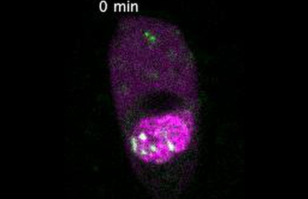
Click image above to watch Movie 1: ASY1 and ZYP1b were tagged by RFP and GFP respectively. At the onset of synaptonemal complex formation, ZYP1b:GFP accumulates in short stretches and progressively elongates into long filaments. It was fully installed when ASY:RFP was no longer observable. [The movie is adapted from (Hu and Prusicki et al., 2026)”]
We also visualized the formation of two daughter cells after cytokinesis by tagging a SNARE protein (SYP132, SOLUBLE N-ETHYLMALEIMIDE-SENSITIVE FACTOR ATTACHMENT PROTEIN RECEPTOR) and α-tubulin (TUA5) with GFP and RFP respectively (Movie 2).
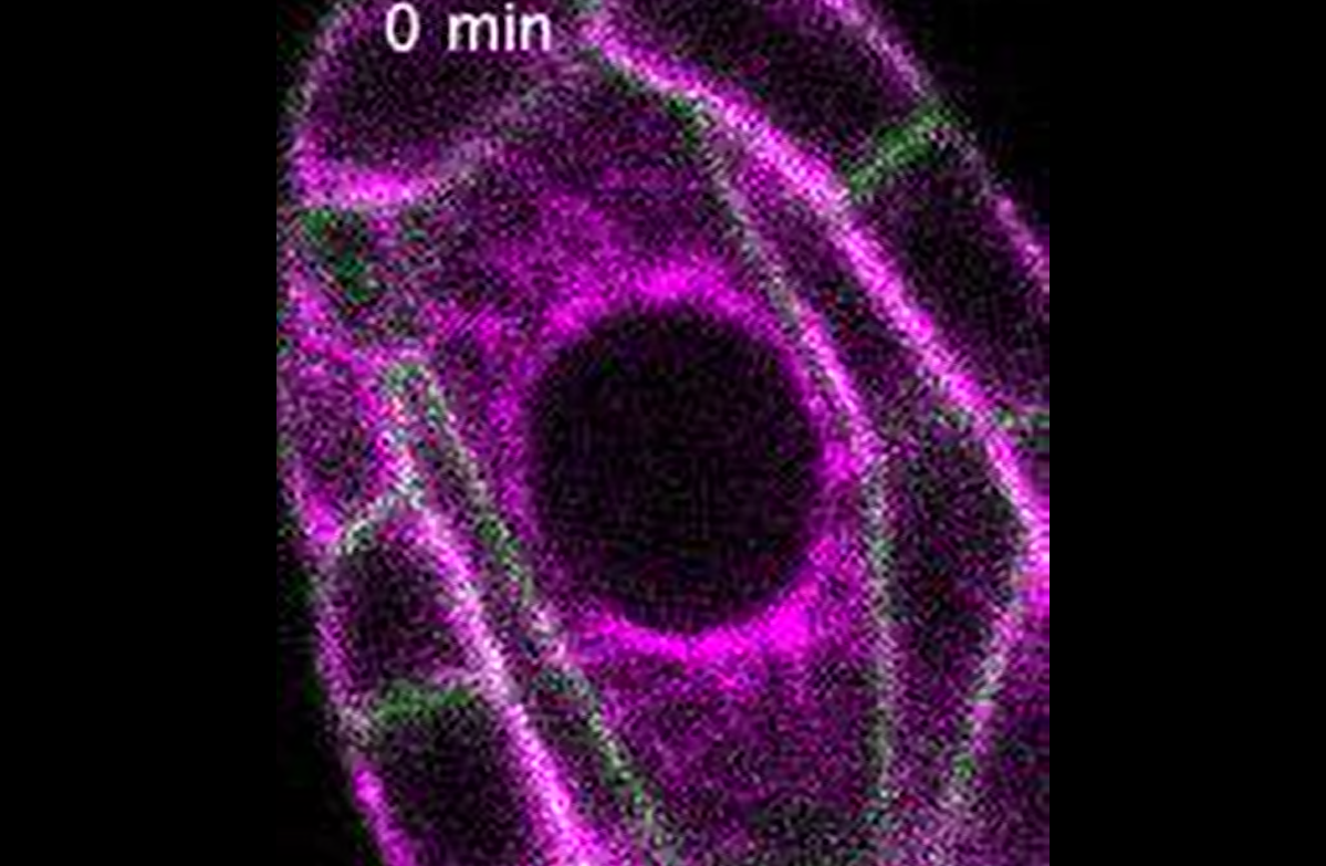
Click image above to watch Movie 2: SYP132 and TUA5 were tagged by GFP and RFP respectively to visualize the formation of two daughter cells after cytokinesis. [The movie is adapted from (Hu and Prusicki et al., 2026)”]
Using this system, we have then analyzed mutants in cyclin-dependent kinase inhibitors in which a designated female meiocyte undergoes several mitotic divisions before undergoing meiosis. This work indicates that meiotic fate is fixed very early in a designated meiocyte, but meiotic structures are flexible and compatible with a mitotic division.
Next steps: With the cytological framework established here, mutants in meiotic regulators can now be analyzed in detail for their defects in the female. Moreover, the effects of environmental conditions on female meiosis, such as heat stress, can now be studied with great temporal resolution.
Original paper: Hu, B., Prusicki, M. A., Stahlmann, K., Wang, Y., & Schnittger, A. (2026). A cytological framework of female meiosis in Arabidopsis. The Plant Cell, koag113. https://doi.org/10.1093/plcell/koag113
Written by: Arp Schnittger (Bluesky: @schnittger-lab.bsky.social)
Edited by: Yee-Shan Ku (LinkedIn: @Yee-Shan Ku)