Review: Integrating cellular electron microscopy with multimodal data to explore biology across space and time
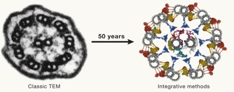 Fifty years ago (1974), Albert Claude, Christian de Duve, and George Palade were awarded the Nobel Prize for their discoveries on the structural and functional organization of the cell, which Claude eloquently framed by writing, “We have entered the cell, the mansion of our birth, and started the inventory of our acquired wealth.” In the subsequent fifty years, amazing new tools and technologies have greatly enhanced our understanding of our cellular inventory. McCafferty et al. have skillfully crafted a comprehensive overview of these methods, beautifully weaving together how they complement each other to provide extraordinary insights into cell structures and compositions in space and time. I particularly enjoyed how the authors melded multiple imaging technologies together along with computational and modeling approaches. As one of several examples, they show using Chlamydomonas how fluorescent microscopy can be combined with ultrastructure expansion microscopy, soft X-ray tomography, cross-linking and co-expression mass spectrometry, single-particle analysis, alpha fold structure prediction, proximity labeling and molecular dynamics modeling. This is a fascinating and inspiring article that makes me eager to see where the next 50 years will take us. (Summary by Mary Williams @PlantTeaching) Cell 10.1016/j.cell.2024.01.005


