Cells are larger than life when ExPOSEd
Plant Science Research WeeklyCells are as small as life gets, but can be much larger than they appear if given room for expansion. This is possible with expansion microscopy, a technique that enables three dimensional cell imaging by physically expanding cellular components for visualization. Although expansion microscopy has been…
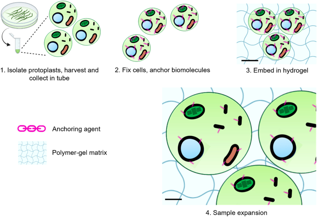
Expanding the resolution limits of conventional microscopy in whole plant tissues
Plant Science Research WeeklyHow can we precisely image plant tissues in super-resolution when approaching the optical limits of conventional microscopes? One solution lies in expansion microscopy, a technique that embeds tissue samples in an expandable hydrogel that proportionally increases the distances between structures, allowing…
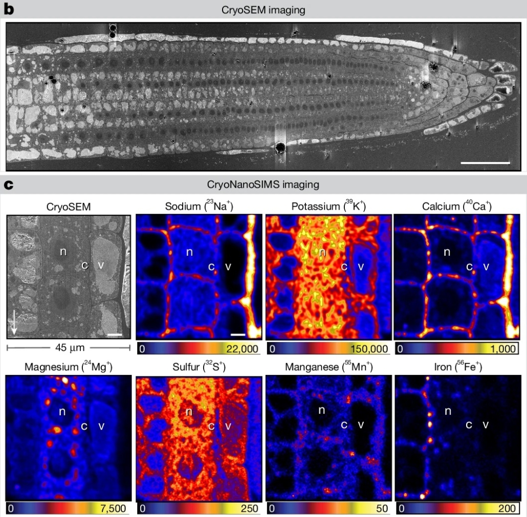
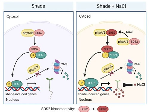
SOS1, salt, and cryo-imaging of subcellular element distribution
Blog, Plant Science Research WeeklyFor living organisms, proper control of element location is just as important as the control of enzyme location, but harder to study. A new study by Ramakrishna et al. uses an exciting new technology, cryo nanoscale secondary ion mass spectrometry ion microprobe, to investigate elemental distribution…
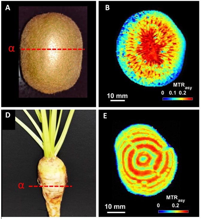
Metabolites through the looking glass with CEST MRI
Plant Science Research WeeklyNon-invasive imaging technologies like computed tomography and magnetic resonance imaging (MRI) have revolutionized medicine by improving diagnostics and guiding treatment. Due to its versatility, MRI also holds potential for plant sciences, where it can be used to visualize and quantify metabolites…
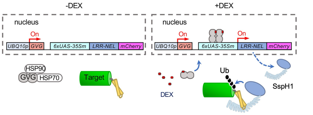
An inducible protein degradation system for rapid depletion of target proteins
Plant Science Research WeeklyTargeting a given protein for degradation at a specific time can be very useful when investigating its function in the cellular context. In this paper, Huang et al. present an inducible protein degradation system tailored for plants, named E3-DART. The system is based on the specific interaction between…
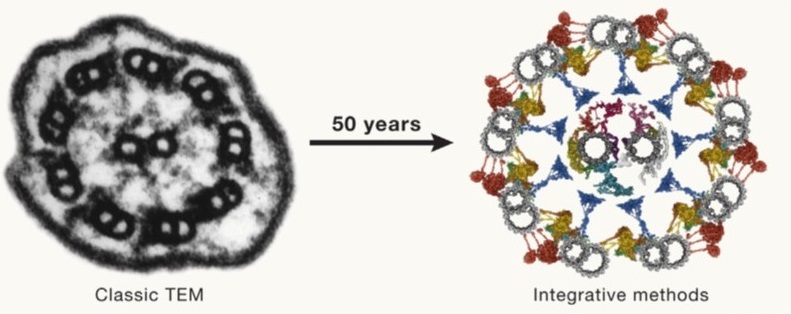
Review: Integrating cellular electron microscopy with multimodal data to explore biology across space and time
Plant Science Research WeeklyFifty years ago (1974), Albert Claude, Christian de Duve, and George Palade were awarded the Nobel Prize for their discoveries on the structural and functional organization of the cell, which Claude eloquently framed by writing, “We have entered the cell, the mansion of our birth, and started the inventory…
Special issue: Human-machine collaboration in plant biology
Plant Science Research WeeklyThis is an excellent article to wrap up this year and lead us into the future. Introducing a special issue of Plant Cell Physiology, Nakajima et al. summarize an exciting collection of papers that look at diverse ways that plant biology can be enhanced through “human-machine collaborations”. Some…
Precise integration of large DNA sequences in plant genomes using PrimeRoot editors
Plant Science Research WeeklyAlthough CRISPR/Cas9 tools have provided new opportunities for genome editing, using these systems to introduce large pieces of DNA has been challenging. A new genome editing technique, “PrimeRoot” (Prime editing-mediated Recombination Of Opportune Targets), was introduced by Sun et al. and shown…
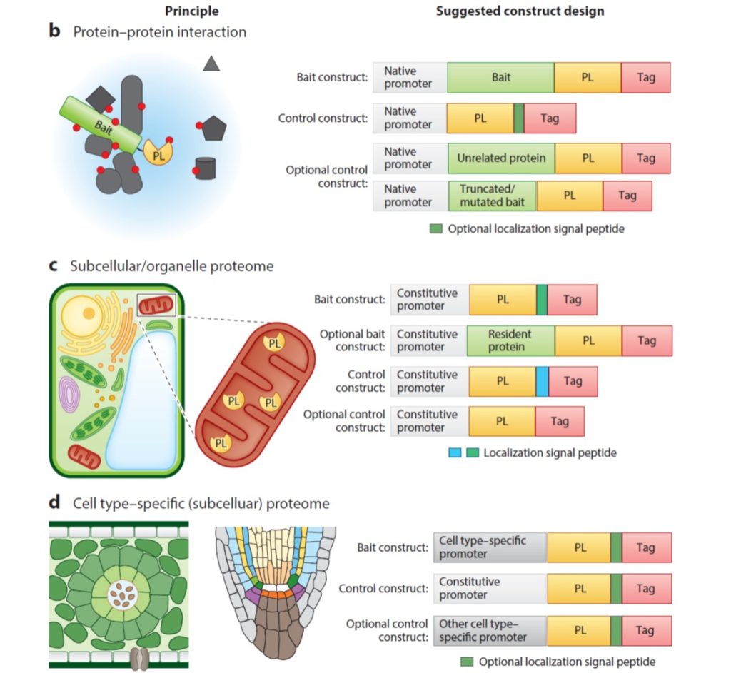
Review: Proximity labeling in plants
Plant Science Research WeeklyGenetic studies can suggest that two proteins function in the same pathway, but how can we figure out if they share the same space? In this review, Xu et al. provide an overview of proximity labeling, a method to identify proteins that co-localize in space. Proximity labeling uses a biotin ligase which…