Review: 3D electron microscopy for imaging organelles in plants and algae (Plant Physiol)
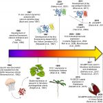
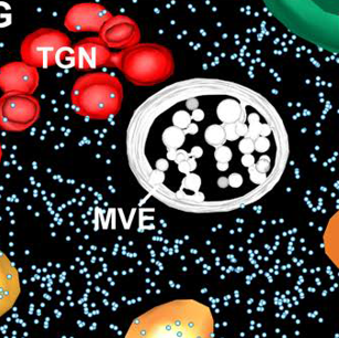 Have you noticed that the quality and resolution of cell images has been getting better and better? Weiner et al. review the recent advances in 3D electron microscopy (EM) technologies that have provided these strikingly beautiful and informative images. Like “classic” transmission EM, EM tomography requires the production of thin sections, either through cryo-preservation (Cryo-EM) or embedding in plastic resin. The sections are imaged from multiple angles by tilting the sample under the electron beam, and then 3D reconstructions are calculated. An alternative approach is serial block-face scanning EM (SBF-SEM). Here, an embedded sample is imaged by scanning EM, then an extremely thin layer removed by an ultramicrotome, and the sample is imaged again. Alternatively, the thin layer is removed by milling with a focused ion beam (FIB-SEM). In correlative light and EM (CLEM), EM methods are combined with light-based imaging, for example fluorescence. This review describes sample preparation required for each of these methods and provides examples of how they have been used in plant cell biology. (Summary by Mary Williams @PlantTeaching) Plant Physiol. 10.1093/plphys/kiab449