CYCLOIDEA-like genes control multiple floral traits

Yang, Wang, Liu, et al. reveal that CYCLOIDEA-like genes control the genetic correlation of floral symmetry, floral orientation, and nectar guide patterns.

https://doi.org/10.1093/plcell/koad115

By Xia Yang1,2 and Yin-Zheng Wang1,2,3
Institutions:
1 State Key Laboratory of Systematic and Evolutionary Botany, Institute of Botany,
Chinese Academy of Sciences, Xiangshan, Beijing 100093, China
2 China National Botanical Garden, Beijing 100093, China
3 College of Life Sciences, University of Chinese Academy of Sciences, Beijing 100049,
China

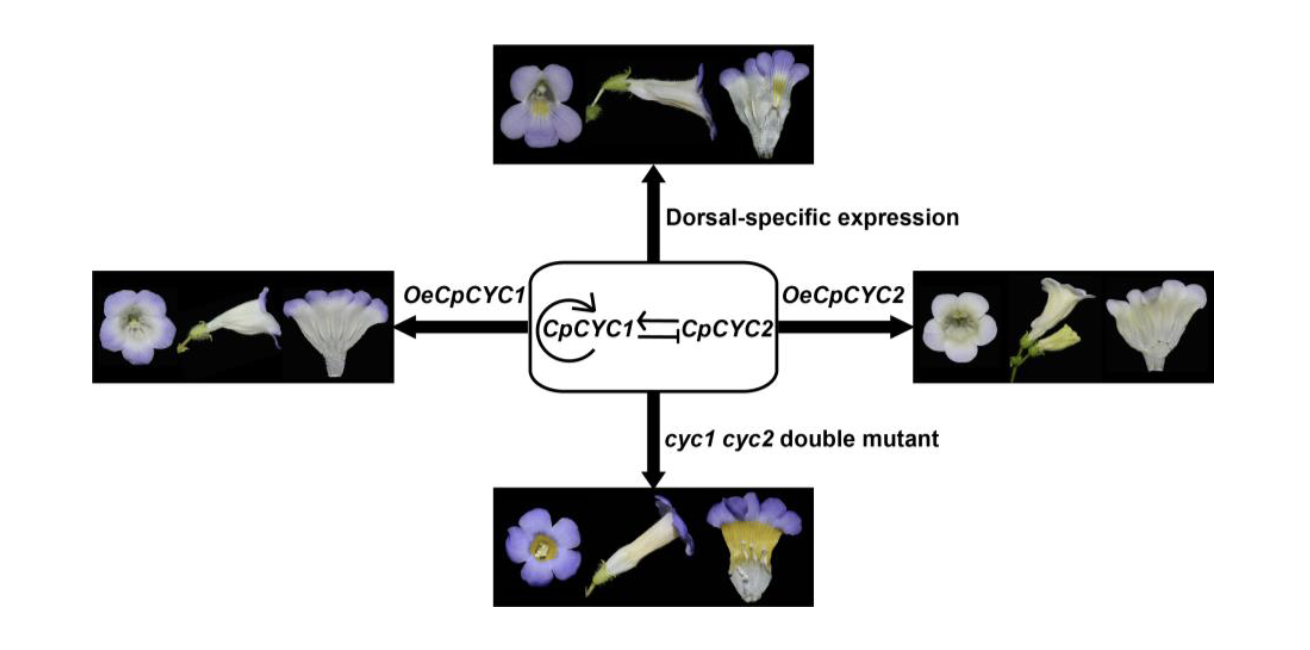
Background: Three types of floral symmetry can be distinguished based on the number of symmetry planes: polysymmetry (with several symmetry planes), monosymmetry (with only one symmetry plane), and asymmetry. Early angiosperms have polysymmetric floral organs, while monosymmetry originated many times from polysymmetry, and several large clades produce predominantly or entirely monosymmetric flowers. In addition to having differential morphologies and sizes in the second and third whorls of floral organs, monosymmetric flowers usually possess horizontal orientation and asymmetric nectar guides. CYCLOIDEA (CYC)-like TCP transcription factors control floral monosymmetry in many species, but little is known about how horizontal orientation and asymmetric nectar guides are achieved.

Question: Are floral symmetry, floral orientation, and nectar guide patterning correlated traits, and are they determined by the same set of master regulators, such as CYC-like genes?

Findings: We selected Chirita pumila (Gesneriaceae) as a model system to address this issue. Plants overexpressing CpCYC1 and CpCYC2 generated dorsalized flowers, with a change in floral orientation from horizontal to upward and the loss of yellow nectar guides in the ventral corolla tube. By contrast, the cyc1 cyc2 double mutant produced ventralized flowers with upward orientation and uniform yellow nectar guides. Therefore, CpCYC1 and CpCYC2 not only determine floral symmetry, but they also regulate floral orientation and nectar guide patterning. CpCYC1 positively regulates itself and down-regulates CpCYC2, while CpCYC2 up-regulates CpCYC1. We also identified the flavonoid biosynthesis-related gene CpF3’5’H, which regulates yellow nectar guide formation. CpCYC1 and CpCYC2 repress yellow pigment formation in nectar guides out the ventral region of the flower, likely by directly repressing CpF3’5’H.

Next steps: Further studies are needed to elucidate how CpCYC1 and CpCYC2 control these distinct floral traits by regulating different target genes or interacting with different co-factors.

Xia Yang, Yang Wang, Tian-Xia Liu, Qi Liu, Jing Liu, Tian-Feng Lü, Rui-Xue Yang, Feng-Xian Guo and Yin-Zheng Wang. (2023). CYCLOIDEA-like genes control floral symmetry, floral orientation, and nectar guide patterning. https://doi.org/10.1093/plcell/koad115

Proximity labeling to examine viral replication complexes

Q. Zhang, Z. Wen, and X. Zhang, et al. use proximity labeling with the TurboID system to identify proteins that are important for replication in plant viruses.

https://doi.org/10.1093/plcell/koad146

Yongliang Zhang1, Xiaofeng Wang2

1 State Key Laboratory of Plant Environmental Resilience and Ministry of Agriculture Key Laboratory of Soil Microbiology, College of Biological Sciences, China Agricultural University, Beijing 100193, China

2 School of Plant and Environmental Sciences, Virginia Tech, Blacksburg, Virginia 24061, USA

 Background:

Upon entry into host cells, positive-strand RNA viruses hijack host factors to remodel specific organelle membranes to form viral replication complexes (VRCs). VRCs provide an optimal microenvironment for protecting and generating progeny viral RNAs. Identifying host components of VRCs is crucial for understanding viral replication mechanisms and developing virus-resistant plants. However, VRCs are membrane associated and dynamic, making it technically challenging to study VRC components using traditional methods for examining protein–protein interactions in plant cells. The recently emerged TurboID-based proximity labeling (PL) approach has become a powerful tool for examining weak, transient, and dynamic molecular interactions in planta. However, TurboID-based PL has not been employed to investigate VRCs in plants.

Question:

Can TurboID-based PL be used to identify key components and interaction networks of VRCs in plants?

Findings:

Using Beet black scorch virus (BBSV) as a model, we systematically investigated the constituents of VRCs by fusing the TurboID enzyme to the BBSV replication protein p23. We identified 185 p23-proximal proteins, including several proteins that are known to interact with p23 or to be critical for BBSV replication. The reticulon family proteins were repeatedly identified. We further demonstrated that reticulon-like protein B2 (RTNLB2) interacts with p23 and plays a pro-viral role in BBSV replication by facilitating the establishment of VRCs. Moreover, RTNLB2 is crucial for replication of several other plant viruses that replicate on endoplasmic reticulum membranes.

Next steps:

Whether RTNLB2 is involved in other aspects of virus infection, such as movement, remains to be investigated. Are other p23-proximal proteins involved in replications of BBSV and/or other plant viruses, if so, what are the underlying mechanisms?

Reference:

Qianshen Zhang, Zhiyan Wen, Xin Zhang, Jiajie She, Xiaoling Wang, Zongyu Gao, Ruiqi Wang, Xiaofei Zhao, Zhen Su, Zhen Li, Dawei Li, Xiaofeng Wang, Yongliang Zhang (2023). RETICULON-LIKE PROTEIN B2 is a pro-viral factor coopted for the biogenesis of viral replication organelles in plants. https://doi.org/10.1093/plcell/koad146

NAC-CYP Mediates Soybean Nodule Senescence

Yu et al. show that activation of the expression of cysteine protease genes by GmNAC039 and GmNAC018 is required for soybean nodule senescence The Plant Cell (2023).

https://doi.org/10.1093/plcell/koad129

By H Yu and Y Cao

Background: Soybean (Glycine max) roots have nodules in which symbiotic bacteria reduce nitrogen gas into ammonium, which nourishes the plants. The termination of symbiotic nitrogen fixation, termed nodule senescence, is an active process and soybean nodules are usually severely senescent when the plant begins flowering, which appears counterproductive, as seed maturation requires a large amount of nitrogen. While much has been discovered about symbiotic signaling and nodule organogenesis over the past two decades, knowledge about nodule senescence is largely limited to decreased nitrogenase activity and leghemoglobin content, physiological changes, and gene expression profiles of senescent nodules. One molecular marker of senescent nodules is an increase in the transcript levels of genes encoding cysteine proteases that break down proteins and degrade nodule cells. However, how nodule senescence is activated at the molecular level and what components activate the expression of senescence-related genes are largely unknown.

Question: What are the key components regulating nodule senescence in soybean?

Findings: We identified two NAC transcription factors from soybean, GmNAC039 and its paralog GmNAC018, as regulators of nodule senescence. GmNAC039 directly binds to the promoters of at least four cysteine protease genes and activates their expression. Knocking out these four cysteine protease genes in soybean delayed nodule senescence and increased nitrogenase activity. Thus, GmNAC039 and GmNAC018 activate the expression of cysteine protease genes to promote nodule senescence in soybean.

Next steps: Nodule senescence is associated with extensive cell degradation, which leads to programmed cell death in nodules. The expression of genes encoding proteins with proteolytic activities is required for nodule senescence. Given that cysteine proteases have key roles in soybean nodule senescence, identifying their target proteins is an important future research goal.

Reference:

Haixiang Yu, Aifang Xiao, Jiashan Wu, Haoxing Li, Yan Duan, Qingshan Chen, Hui Zhu, and Yangrong Cao. (2023) GmNAC039 and GmNAC018 activate the expression of cysteine protease genes to promote soybean nodule senescence https://doi.org/10.1093/plcell/koad129

Turnover of Complex I is regulated by FTSH3 that recognises the PSST subunit

Ghifari et al. explore the mechanisms of Complex I disassembly and turnover.

https://doi.org/10.1093/plcell/koad128

Abi S. Ghifari and Monika W Murcha

School of Molecular Sciences, The University of Western Australia

Background: Oxidative phosphorylation (OXPHOS) is the central process of aerobic respiration in plant mitochondria. Complex I, the first entry point and largest complex of the OXPHOS pathway, begins the OXPHOS process by oxidizing the high-energy intermediate NADH and transferring electrons to the mobile electron carrier ubiquinone. High redox activity and constant exposure to reactive oxygen species render Complex I subunits prone to oxidative damage, resulting in a high turnover rate. Despite recent advances in the structural determination of plant Complex I, how Complex I degradation and turnover is regulated remains enigmatic.

Question: Which factors determine Complex I turnover, and how is this process mechanistically regulated?

Findings: Using two independent Arabidopsis thaliana EMS mutants generated in a Complex I defective background, we show that FTSH3, a mitochondrial matrix-facing inner membrane-bound protease, facilitates the unfolding of Complex I matrix arm subunits for degradation and turnover. For this function, FTSH3 interacts directly with PSST, a Complex I matrix arm domain subunit. This interaction is mediated by the ATPase domain of FTSH3 and the N-terminal domain of PSST. Mutations in these domains prevent the interaction between these two factors, slowing the turnover rate of matrix arm subunits, resulting in enhanced Complex I subunit abundance and activity.

Next steps: We plan to assess the specificity of FTSH3 in regulating the turnover of other OXPHOS complexes, as well as its modulation of the activities of other mitochondrial proteases. By identifying the role of FTSH3 with regards to other OXPHOS subunits and proteases, we can provide a more comprehensive view of how of mitochondrial OXPHOS protein turnover is regulated.

Reference:

Abi S. Ghifari, Aneta Ivanova, Oliver Berkowitz, James Whelan, Monika W. Murcha. (2023). FTSH PROTEASE 3 facilitates complex I degradation through a direct interaction with the complex I subunit PSST https://doi.org/10.1093/plcell/koad128

Enhanced discovery of plant RNA-binding proteins

Zhang, Zu, et al. develop a method to identify RNA-binding proteins that bind poly(A) and non-poly(A) RNAs.

Yong Zhang and Devinder Sandhu; University of California Riverside and US Salinity Laboratory (USDA-ARS), Riverside, CA, USA

Background: RNA-binding proteins (RBPs) are important for controlling the fate of cells and for regulating important processes during development. Scientists study RBPs in a systematic way to understand how they control genes. However, studying RBPs in plants is difficult because plants have complex tissues. Additionally, most plant RBPs have only been discovered by studying a small fraction of RNA molecules called polyadenylated (poly(A)) RNA, which may not give a complete picture of all the RBPs. As a result, the true number of RBPs in plants may be much higher than previously thought.

Question: How can we develop a method to capture all the RBPs in plants, including those that bind to different types of RNA, i.e. poly(A) and non-poly(A), to get a comprehensive and unbiased understanding of RBPs in plants?

Findings: We developed a robust plant phase extraction (PPE) method and identified both constitutive and tissue-specific RBPs from Arabidopsis leaf and root tissues under normal growth conditions and salinity stress. We identified several previously un-annotated RBPs, some of which are involved plant responses to salt stress. More importantly, 40% of the RBPs discovered by PPE are non-poly(A) RBPs. Interestingly, many of the new RBPs are metabolic/catabolic enzymes that do not have the classic RNA-binding domains that scientists typically associate with RBPs. We also uncovered 38 domains from these enzymes as putative RNA-binding domains and provided evidence that some catalytic domains of these enzymes can directly bind RNA.

Next steps: It would be intriguing to study how these salt-responsive RBPs respond to salinity stress at the post-transcriptional level and how RNA competes or cooperates with the substrate for these metabolic/catabolic enzymes in the regulation of RNA processing and intermediary metabolism.

Reference:

Yong Zhang, Ye Xu, Todd H. Skaggs, Jorge F.S. Ferreira, Xuemei Chen and Devinder Sandhu (2023) Plant phase extraction: A method for enhanced discovery of the RNA-binding proteome and its dynamics in plants https://doi.org/10.1093/plcell/koad124

The catalytic domain of cellulose synthase: More than just cellulose biosynthesis

Huang et al. demonstrate that the conserved catalytic domain of cellulose synthase 6 (CESA6) is involved in trafficking, protein dynamics, and complex formation. The Plant Cell (2023)

https://doi.org/10.1093/plcell/koad110

Lei Huang,1,2 Weiwei Zhang,1,3 Xiaohui Li,1,2 and Christopher J. Staiger1,2,3

1Department of Botany and Plant Pathology, Purdue University, West Lafayette, IN, 47907, USA

2Center for Plant Biology, College of Agriculture, Purdue University, West Lafayette, IN, 47907, USA

3Department of Biological Sciences, Purdue University, West Lafayette, IN, 47907, USA

 Background: The cell wall plays a vital role in the survival and growth of plants, and provides abundant renewable biopolymers, like cellulose, that have significant economic importance. Cellulose biosynthesis occurs at the cell surface through a plasma membrane (PM)-localized multimeric protein complex named the cellulose synthase (CESA) complex (CSC). CESAs are synthesized in the endoplasmic reticulum, transported to the Golgi for CSC complex assembly, and delivered to the PM through secretory vesicles. Recently, we identified a small molecule inhibitor, Endosidin 20, that targets the catalytic domain of CESA6 and apparently affects CSC trafficking to the PM.

 Question: The large catalytic domain of plant CESA contains several key motifs that are also present in bacterial CESAs. How do these conserved motifs and the catalytic domain of plant CESAs contribute to CESA protein dynamics, complex formation and/or trafficking?

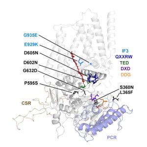
Findings: We created and tested 18 single amino acid replacement mutations in the CESA6 catalytic domain. Using quantitative live-cell imaging of the dynamics of CESA6 labeled with the yellow fluorescent protein (YFP-CESA6), we found that multiple mutations in the DXD, TED and QXXRW motifs, but not the DDG motif, caused reduced motility of PM-localized CSCs, indicative of reduced cellulose biosynthesis activity. Additionally, various mutations affected Golgi-to-PM trafficking of the CSC. Using principal component analysis of nine biochemical and cellular parameters, the 18 mutations separated into two major groups. Group I mutations caused CSC trafficking defects whereas Group II mutations, especially in the QXXRW motif, affected CESA protein folding and/or CSC rosette formation in the ER or Golgi with consequences for post-Golgi trafficking.

 Next steps: CESA-associated proteins participate in plant tolerance to salt and drought stresses, CSC exocytosis, etc. through interactions with the catalytic domain of CESA. It will be interesting to test how our collection of point mutations in CESA6 affect plant response to adverse stresses and/or modulate interactions with CESA-associated proteins.

Reference:

Huang L., Zhang W., Li X., Staiger C.J., Zhang C. (2023). Point mutations in the catalytic domain disrupt cellulose synthase (CESA6) vesicle trafficking and protein dynamics. https://doi.org/10.1093/plcell/koad110

Review: Red macroalgae in the genomic era

I highly recommend this excellent and accessible article by Borg et al. that provides an overview of the red macroalgae, which “may have been the first eukaryotic lineage to have evolved complex multicellularity”. It’s full of fascinating information: although 97% of red algal species are marine, one lives in sloth hairs, and nori (sushi seaweed) is made from red algae (Porphyra and Pyropia genera). This article also gives an excellent and fascinating overview of red algal cell biology; from their unique cell wall polysaccharides that provide them with strength and elasticity (and us with agarose) to their unusual and highly efficient light-harvesting antennae (phycobilisomes) and their lack of plasmodesmata. There’s a tremendous diversity and variation in the reproductive biology of the red algae spanning from triphasic life cycles to the presence of trichogynes that receive the male nucleus for fertilization; interestingly, the authors point out that this great diversity offers the opportunity to explore questions about the evolutionary and ecological importance of sex. As the title suggests, genomic tools and the emergence of some model species are now shedding further light on these fascinating and important organisms. Summary by Mary Williams @PlantTeaching) New Phytol. 10.1111/nph.19211

Review: How plant roots respond to waterlogging

As the hottest year on record, 2023 has truly been a global annus horribilis during which humans, other animals, and domesticated and wild plants have faced unprecedented environmental challenges. In the past month alone, torrential rainfalls have wreaked havoc in Asia, the Middle East, and many parts of Africa, Europe, and the Americas. Frightenedly, disruptive flooding is expected to get worse, adding to food security concerns. Therefore, it’s timely to read about how plants respond to waterlogging in the new review by Daniel and Hartman. The main challenge in waterlogging is hypoxia, oxygen deprivation. The plant senses its waterlogged state through both hypoxia and an accumulation of ethylene, which is not able to diffuse away under water. Some plants have adaptations that enable them to tolerate at least some waterlogging, such as aerenchyma (air channels in roots), the ability to enter a quiescent state, or rapid elongation to reach above the water level. However, for non-adapted plants, even short periods can be lethal. The review covers plant responses from sensing, metabolic changes, and growth responses including changes to root system architecture and gravitropic setpoints (plants are amazing). It also discusses the very important question of translating from simple experimental systems (e.g., Arabidopsis on Petri plates) to real-world conditions, and makes the plea for moving towards “less artificial” experimental conditions. (Summary by Mary Williams @PlantTeaching) J. Exp. Bot. 10.1093/jxb/erad332

Plant Physiology Focus Issue: Plant Cell Polarity

The September issue of Plant Physiology has a focus on plant cell polarity, which plays a pivotal role in the fundamental processes that dictate plant growth, development, and adaptation. By establishing distinct regions within cells, plant cell polarity is crucial for regulating asymmetric cell divisions, guiding the direction of cell expansion, and determining the spatial distribution of essential cellular components. Understanding the mechanisms and regulatory pathways involved in plant cell polarity holds immense promise for enhancing crop productivity, optimizing plant responses to changing environments, and ultimately contributing to sustainable agriculture and ecological resilience. Don’t miss the editorial overview by Dong et al., and keep your eyes open for the registration link for a Focus Issue webinar to be held Sept. 25.  (Summary by Mary Williams @PlantTeaching) Plant Physiol. 10.1093/plphys/kiad436.