Selective Chloroplast Microautophagy
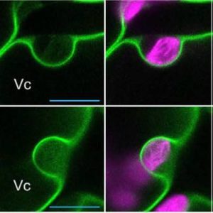 When plants are exposed to excessive light, photoinhibition occurs and chloroplasts become damaged. Photodamaged chloroplasts undergo vacuolar digestion through a poorly understood autophagic process called chlorophagy. In general, cell biologists recognize two types of autophagy: macroautophagy and microphagy. Macroautophagy is a form of autophagy in which a membrane forms around the material to be digested before it fuses with the vacuole or lysosome. Microautophagy is a distinct form of autophagy in which the material to be digested fuses directly with the vacuole or lysosome. These considerations raise the question of whether chlorophagy is a type of microphagy or macrophagy? Nakamura et al. (10.1104/pp.18.00444) report upon their detailed observations of chlorophagy in Arabidopsis (Arabidopsis thaliana) mesophyll cells damaged by high light intensities. They provide evidence that photoinhibition produces abnormally swollen chloroplasts. These damaged chloroplasts are labeled by autophagosome structures for selective elimination via chlorophagy, thereby avoiding the cytoplasmic accumulation of dysfunctional chloroplasts. This selective removal is triggered by an imbalance of membrane potential across the chloroplast envelope resulting from membrane damage. Cytological evidence is presented that suggests that chlorophagy is achieved directly by the tonoplast-mediated sequestering of swollen chloroplasts and, thus, represents a microautophagy-type process.


