Review. Imaging flowers: a guide to current microscopy and tomography techniques to study flower development (J. Exp. Bot.)
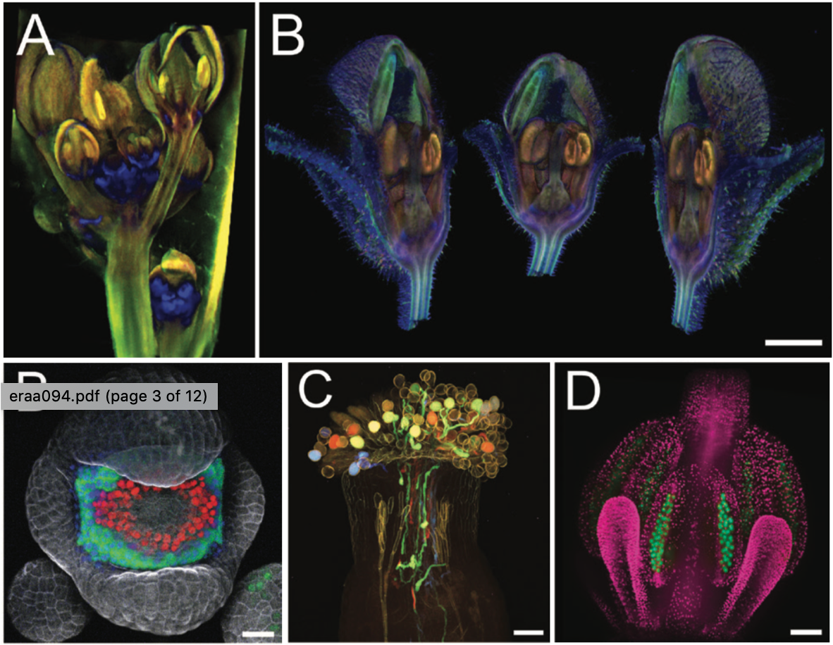 Flowers bear the reproductive organs and determine the reproductive success of plants by producing fruits and seeds. Flowers usually include four whorls of organs: sepals, petals, stamen and carpel. In this review, Prunet and Duncan discuss various microscopic and tomographic techniques to image flower organs with different levels of resolution and at different depths. They focus on the point-scanning confocal, spinning disc confocal and two-photon microscopy and their pros and cons, including the lateral and axial resolution, imaging depth, live imaging and fluorescent protein compatibility. The authors also describe high-resolution microscopy techniques like structured illumination microscopy (SIM), stimulated emission depletion microscopy (STED), light sheet microscopy, and single molecule localization microscopy (SMLM) techniques which give high resolution images at the cost of speed. Optical microscopy cannot provide accurate 3D deep tissue images, but this problem can be circumvented by tomography. Tomography techniques include optical projection tomography (OPT), macro-OPT, X-ray microscopy (XRM), X-ray tomography (XRT). These methods acquire 3D images at a fast speed but at the cost of cellular resolution. Finally, the authors discuss the various reporters and sensors developed recently for better quantification of gene activity and hormone signaling, and the software to analyze the huge datasets created using these techniques. (Summary by Vijaya Battula @Vijaya_Batthula). J. Exp. Bot. 10.1093/jxb/eraa094


")