Nanoscale movements of cellulose microfibrils in primary cell walls ($)
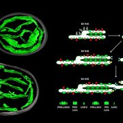
 Cell walls are complex mixtures of cellulose microfibrils, proteins and other materials. Their mechanical properties can be measured and modeled, but it is not always simple to translate these measurements to changes at the molecular level. Zhang et al. used atomic force microscopy to provide an unprecedented view of how cellulose microfibrils respond to stress and strain (if you are a bit fuzzy on the difference between stress and strain see Baskin’s excellent News and Views ). The authors imaged the microfibrils while the cell wall was subjected to plastic and elastic extension and stress relaxation by the addition of a cell-wall loosening enzyme. A key finding is that in some conditions the microfibrils are able to slip relative to the surrounding matrix. The authors also describe new insights into microfibril connections and anchoring. Nature Plants 10.1038/nplants.2017.56


Leave a Reply
Want to join the discussion?Feel free to contribute!