Real-time conversion of tissue-scale mechanical forces into an interdigitated growth pattern (Nature Plants)
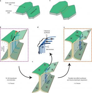 In many plants, epidermal pavement cells lock together through interdigitating lobes, much like the pieces of a jigsaw puzzle. These cells start out as regular squarish cells with straight walls, which then bow out and curve. Here, Belteton et al. investigate how lobes form in cotyledon pavement cells. Using a 3D long-term time-lapse imaging pipeline, they imaged expanding leaves, labeling plasma membranes, microtubules and plasmodesmata; these data allowed them to quantify very localized rates of wall growth in 3D. This pipeline was also used to image cells grown in the presence of various inhibitors as well as cells from mutants associated with wall or cytoskeleton defects. Finite-element modeling of these data simulated stress-strain relationships in the walls. These studies identified a critical role in lobe formation of microtubules that are transfacial: i.e., they span both periclinal (parallel to the leaf surface) and anticlinal (perpendicular to the leaf surface) surfaces. These localized microtubule arrays pattern cellulose microfibrils, causing localized asymmetry which leads to lobe formation. These transfacial microtubles can coordinate the growth rate of upper and lower periclinal walls, allowing the anticlinal wall to maintain a constant tilt angle. This mechanistic model of lobe formation provides a foundation to analyse the cellular basis of leaf morphology and function. (Summary by Mary Williams @PlantTeaching) Nature Plants 10.1038/s41477-021-00931-z


