Another Step Closer to Understanding Plant Cell Wall Biosynthesis: The Crystal Structure of FUCOSYLTRANSFERASE1[

IN BRIEF by Nancy R. Hofmann  [email protected]

Plant cell walls consist of cellulose microfibrils embedded in a matrix of polymers including hemicelluloses. As one of the main hemicelluloses in the cell walls of dicots, xyloglucan is an important target of study to understand plant cell walls in general and for polymer applications in biotechnology. Xyloglucan consists of a β-1,4-linked backbone of glucosyl residues with or without side chains, the most common of which is a xylosyl residue. The side chains themselves can harbor further side chains, and, ultimately, 24 different side chains can make up xyloglucan. Biosynthesis of xyloglucan (reviewed in Pauly and Keegstra, 2016) involves members of the glycosyltransferase (GT) superfamily, including a fucosyltransferase, which was the first plant cell wall-biosynthetic enzyme discovered. Now, in a Breakthrough Report from Rocha et al. (2016), Arabidopsis thaliana FUCOSYLTRANSFERASE1 (FUT1) provides the first crystal structure of a plant cell wall biosynthesis enzyme.

FUT1 transfers fucose from the donor GDP-fucose to galatosyl residues on xyloglucan and is a member of glycosyltransferase family 37 (GT37). Rocha and coworkers solved the crystal structures of the soluble portion of FUT1 both in the apo form and bound to GDP and a xyloglucan oligosaccharide acceptor. The most similar known structures were those of fucosyltransferases from other GT families, including Caenorhabditis elegans POFUT1 and human POFUT2, which mediate O-fucosylation of proteins in those species. These related structures have a GT-B fold, which canonically includes two α/β/α Rossmann-fold domains, termed the N- and C-domains, with the active site in a cleft between them. The FUT1 structure represents a GT-B variant not previously observed (see figure). Instead of the α/β/α N-domain, the FUT1 N-domain has a β-sheet with α-helices on one side, but loops on the other. The C-domain has a more typical Rossmann fold, but an extra C-terminal domain forms β-hairpins that lie against the N-domain. This extra domain contacts both the N- and C-domains and forms part of the acceptor binding site.

fucosyltransferase

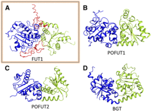
FUT1 adopts a variant of the GT-B superfamily fold. (A) FUT1 structure reported in this work. (B) and (C) Two of the most similar reported structures: (B) POFUT1 (PDB code 3ZY6) and (C) POFUT2 (PDB code 4AP5). (D) Canonical GT-B fold of the phage T4-glucosyltransferase (BGT; PDB code 1BGT), with two similar Rossmann domains: the N-domain (blue) and the C-domain (green). (Adapted from Rocha et al. [2016], Supplemental Figure 1.)

The mechanism of GDP binding by FUT1 appears to be quite similar to that in the four most similar structures, despite FUT1 sharing <20% sequence identity with the corresponding proteins. Importantly, the conserved residues involved in binding include those in three motifs (I, II, and III) that were previously reported to be important for GT activity. Thus, the FUT1 structure demonstrates that these residues are necessary for GT activity because of their importance in donor sugar binding.

The authors also use the structure to explain FUT1’s substrate specificity. In contrast to the second galactosyl in the acceptor subunit, the first residue is on the opposite side of the β-1,4-d-glucan backbone and does not make any contact with FUT1. Accordingly, FUT1 fucosylates the second, but not the first, galactosyl residue in a xyloglucan acceptor. The extra C-terminal domain of FUT1 also interacts with acceptor. Interestingly, this domain is present in all 10 Arabidopsis thaliana GT37 family members, among which only FUT1 acts on xyloglucan. Rocha et al. were able to divide this family into three subgroups, based on their sequences in the region that contributes to the anchoring of the xyloglucan subunit in FUT1. Their analysis provides an opening into predictions of substrate specificity based on primary sequence, which has proved quite difficult among GT family members.

Overall, this work adds another first to the rich history of fucosyltransferase research in plants and provides insight into the biosynthesis of an important plant cell wall component as well as into the function of the huge family of glycosyltransferases in general.

REFERENCES

Pauly, M., and Keegstra, K. (2016). Biosynthesis of the plant cell wall matrix polysaccharide xyloglucan. Annu. Rev. Plant Biol. 67: 235–259.

Rocha, J., Cicéron, F., de Sanctis, D., Lelimousin, M., Chazalet, V., Lerouxel, O., and Breton, C. (2016). Structure of Arabidopsis thaliana FUT1 reveals a variant of the GT-B class fold and provides insight into xyloglucan fucosylation. Plant Cell 28: 2352–2364.

It’s Not Easy Not Being Green: Breakthroughs in Chlorophyll Breakdown

chlorophyll-breakdown

IN BRIEF by Jennifer Mach [email protected]

Plants can dispose of organs such as leaves and recycle the nutrients in these organs into new leaves, seeds, or storage organs. However, when separated from its photosystem proteins, chlorophyll can be phototoxic, absorbing light and producing high-energy electrons. The complex chlorophyll degradation pathway solves this problem by breaking down chlorophyll into colorless catabolites that are stored in the vacuole (reviewed in Christ and Hörtensteiner, 2014).

One of the first steps in chlorophyll breakdown is the removal of the Mg from chlorophyll a to form pheophytin a. Pheophytin a also functions as part of the D1/D2 complex in photosystem II, helping to drive electron transfer during photosynthesis. However, the identity (and even existence) of a putative Mg-dechelatase that removes Mg from chlorophyll a has remained unclear. In a recent Breakthrough Report, Shimoda et al. (2016) reason that mutants in Mg-dechelatase would have a stay-green phenotype; therefore, the authors examine proteins related to pea (Pisum sativum) STAY-GREEN (SGR), which causes the green-cotyledon phenotype described by Mendel. The Arabidopsis thaliana genome has three SGRs: SGR1, SGR2, and STAY-GREEN LIKE (SGRL). Recombinant SGR1 and SGR2 expressed in wheat germ extract showed high dechelating activity on chlorophyll a; by contrast, SGRL showed higher activity on chlorophyllide a. Also, expression of SGR1 caused an increase in pheophytin a in transgenic Arabidopsis plants and in cyanobacteria. Arabidopsis mutants for pheophytinase, the next enzyme in the chlorophyll breakdown pathway, also accumulated more pheophytin a when expressing SGR1. Expression of SGR1 in Arabidopsis also caused decreases in chlorophyll contents and in photosystem proteins, but not other chloroplast proteins, indicating that SGR can act on the chlorophyll in protein complexes and thus regulate the amounts of these complexes.

Chlorophyll breakdown initiates in the chloroplast, where enzymes transform pheophorbide a into primary fluorescent chlorophyll catabolite (pFCC). In the cytoplasm, pFCC undergoes species-specific modifications; all species studied also hydroxylate pFCC at the C32 position to form hydroxy-pFCC. Using chromoplasts from bell pepper (Capsicum annuum), Hauenstein et al. (2016) find that the activity that hydroxylates pFCC localizes in the plastid membrane. This activity also requires O2 and ferredoxin, similar to the Rieske-type oxygenase PHEOPHORBIDE a OXYGENASE (PAO). The Arabidopsis genome encodes five Rieske-type oxygenases; of these, three localize to chloroplast membranes: PAO, TRANSLOCON AT THE INNER CHLOROPLAST ENVELOPE55 (TIC55), and PROTOCHLOROPHYLLIDE-DEPENDENT TRANSLOCON AT THE INNER CHLOROPLAST ENVELOPE52 (PTC52). The tic55 and ptc52 mutants showed no visible phenotypes, but the tic55 mutants showed decreased levels of hydroxy-pFCC. By contrast, the ptc52 mutants showed wild-type levels of hydroxy-pFCC. Previous work showed that TIC32 and TIC62 function with TIC55, possibly delivering electrons for the TIC55 redox cycle; however, the authors observed no changes in hydroxylated phyllobilins in tic32 and tic62 mutants.

The finding that TIC55 functions in chlorophyll breakdown proved surprising because previous work implicated TIC55 in protein transport into the chloroplast, acting with TIC32 and TIC62 to sense the redox state of the chloroplast and regulate protein import. However, this finding casts doubt on its potential function in protein transport. Therefore, the roles of these intriguing chloroplast proteins, TIC55 and the SGRs, remain interesting subjects for future work, specifically whether and how SGRs regulate the protein levels of photosystem and light-harvesting complex proteins and whether TIC55 has additional functions in other aspects of chloroplast biology. Given that the tic55 mutants show no obvious visible phenotype, the importance of hydroxylation of phyllobilins also remains an outstanding question.

REFERENCES

Christ, B., and Hörtensteiner, S. (2014). Mechanism and significance of chlorophyll breakdown. J. Plant Growth Regul. 33: 4–20.

Hauenstein, M., Christ, B., Das, A., Aubry, S., and Hörtensteiner, S. (2016). A role for TIC55 as a hydroxylase of phyllobilins, the products of chlorophyll breakdown during plant senescence. Plant Cell 28: 2510–2527.

Shimoda, Y., Ito, H., and Tanaka, A. (2016). Arabidopsis STAY-GREEN, Mendel’s green cotyledon gene, encodes magnesium-dechelatase. Plant Cell 28: 2147–2160.

Field of Genes: Uncovering EGRINs (Environmental Gene Regulatory Influence Networks) in Rice That Function during High-Temperature and Drought Stress

IN BRIEF by Jennifer Lockhart [email protected]

Heat and drought stress greatly restrict crop productivity, but most of what we know about a plant’s response to these stresses comes from controlled laboratory studies. This factor, along with the complex nature of these responses, has hampered efforts to breed and engineer crops with improved stress tolerance. Plants respond to fluctuating environments through the altered expression of numerous genes whose transcription is tightly coordinated. Enter the EGRINs (environmental gene regulatory influence networks), which orchestrate the timing and rate of transcription of these genes in response to environmental signals. Transcription factors (TFs), comprising the core of EGRINs, interact with conserved regulatory elements in genomic DNA to modulate their expression. Since plants can’t run away from their environments, they likely have scores of EGRIN modules that help them cope with changing environments. To date, most EGRINs have been identified using algorithms based on transcriptome data under the assumption that gene expression patterns can be used to predict the relationships between TFs and the genes they regulate. However, simply identifying groups of genes that are coexpressed under stress conditions fails to capture the complexity of these interactions. Since EGRINs affect chromatin availability in multiple regions of the genes they regulate, Sullivan et al. (2014) used changes in chromatin accessibility and the identification of transcription factor footprints, rather than changes in gene expression, to identify putative stress-responsive transcriptional networks in Arabidopsis thaliana.

Wilkins et al. (2016) went one step further by combining chromatin accessibility analysis with coexpression data and network inference algorithms to uncover EGRINs in drought- and heat-stressed rice (Oryza sativa), a major crop whose yields are threatened by climate change. Specifically, the authors investigated the responses of five tropical Asian rice cultivars to high-temperature and water-stress conditions using both hydroponically grown plants in a controlled environment and plants that were naturally exposed to stress conditions in the field (see figure). The authors performed time-series transcriptome analysis (RNA-seq) and chromatin accessibility analysis (ATAC-seq; assay of transposase accessible chromatin) to connect TFs with genes containing accessible, known TF binding motifs in their promoters in order to organize what is currently known about putative regulatory interactions. This analysis uncovered 5447 putative target genes for 445 transcription factors. After various filtering steps, the authors performed network component analysis, a technique used to build upon known networks based on output such as gene expression data (Liao et al., 2003). Final networks were deduced using a network-learning tool known as the Inferelator algorithm (Greenfield et al., 2013).

ergin

This analysis uncovered possible EGRINs involving 4052 target genes regulated by 113 TFs. Most TFs appear to regulate target genes in response to a single stress treatment, whereas a small number of TFs regulate their targets under multiple stress conditions. The use of data from both growth chamber- and field-grown plants allowed the authors to infer EGRINs that function in response to both rapid, controlled stress conditions and fluctuating environments in the field, producing an unusually rich set of data. The EGRINs inferred in this study could be refined and may be used someday to identify high-priority targets for plant breeding and biotechnology programs, fulfilling the dreams of biologists and farmer alike.

REFERENCES

Greenfield, A., Hafemeister, C., and Bonneau, R. (2013). Robust data-driven incorporation of prior knowledge into the inference of dynamic regulatory networks. Bioinformatics 29: 1060–1067.

Liao, J.C., Boscolo, R., Yang, Y.-L., Tran, L.M., Sabatti, C., and Roychowdhury, V.P. (2003). Network component analysis: Reconstruction of regulatory signals in biological systems. Proc. Natl. Acad. Sci. USA 100: 15522–15527.

Sullivan, A.M., et al. (2014). Mapping and dynamics of regulatory DNA and transcription factor networks in A. thaliana. Cell Reports 8: 2015–2030.

Wilkins, O., Hafemeister, C., Plessis, A., Holloway-Phillips, M.-M., Pham, G.M., Nicotra, A.B., Gregorio, G.B., Jagadish, S.V.K., Sptiningsih, E.M., Bonneau, R., and Purugganan, M. (2016). EGRINs (environmental gene regulatory influence networks) in rice that function in the response to water deficit, high temperature, and agricultural environments. Plant Cell 28: 2365–2384.

A Breakthrough in Monocot Transformation Methods

IN BRIEF by Nancy Hofmann [email protected]

The ability to generate transgenic plants without regard to cultivar or genotype can be considered a holy grail of cereal crop transformation. Despite years of effort, it has been remarkably difficult to develop efficient methods for transformation of cereals. The preferred methods generally involve Agrobacterium-mediated transformation of cultured tissue or immature embryos, followed by callus culture to regenerate plants (reviewed in Shrawat and Lörz, 2006). Unfortunately, the capacity of Agrobacterium to infect monocots is limited to a narrow range of genotypes and the utility of the technique is further limited by the recalcitrance of many genotypes to callus formation and regeneration. A Breakthrough Report from Lowe et al. (2016) describes an exciting new approach to boost monocot transformation rates in a broad range of genotypes.

Lowe et al. took advantage of morphogenic genes to create a biological context conducive to transformation efficiency. Their transformation constructs included Zea mays (maize) Baby boom (Bbm) and Wuschel2 (Wus2), homologs of which in Brassica napus and Arabidopsis thaliana promote transition from vegetative to embryonic growth (Boutilier et al., 2002; Zuo et al., 2002). The authors used these constructs for Agrobacterium-mediated transformation of immature embryos from four maize inbred lines. These lines showed markedly higher transformation rates, going from 0-2% in the controls to 25-51% in the presence of Wus and Bbm.

Lowe et al. designed their constructs with flanking LoxP recombination sites and CRE recombinase driven by a drought-inducible promoter. Accordingly, desiccation treatment of the transgenic tissue allowed excision of the Wus, Bbm, and CRE expression cassette. The final transformation efficiency, accounting for all three stages of the process (transformation, excision, regeneration of healthy plants), for the four tested lines ranged from 3-11% (calculated based on the number of initial embryos used and the final number of healthy transgenic plants obtained). This level of recovery of transgenic plants is high enough to be useful for large-scale production both for commercial purposes and in primary research.

Among a panel of 50 maize inbred lines, 33 successfully produced transgenic calli upon transformation with Wus and Bbm. Thus, although not yet fully genotype independent, this approach greatly increases the range of cultivars available for transformation. Importantly, it can be used on a variety of initial tissues. It can be costly and time-consuming to obtain a consistent supply of immature embryos, but Lowe et al. obtained healthy transgenic plants by directly transforming mature seed tissues or seedling leaf segments (see Figure) – skipping the usual explant culture step before transformation and further facilitating large-scale transformation efforts.

picture1

This morphogenic-gene based approach also worked in other monocot crops, increasing transformation frequency from 1.9% to 18% in sorghum and from 0-3% to 15-43% in rice, whereas sugarcane callus showed an almost fantastical increase from 2% to 274/885% (i.e., multiple transgenic events recovered per initial starting tissue). In extending the range of species, genotypes and tissues that can be used for efficient transformation, Lowe et al. have given us a beautiful example of what can be accomplished by combining basic research, technical expertise, and knowledge of practical problems facing mainstream applications.

REFERENCES

Boutilier, K., Ovringa, R., Sharma, V.K., Kieft, H., Ouellet, T., Zhang, L., Hattori, J., Liu, C-M., Van Lammeren, A.A.M., Miki, B.L.A., Custers, J.B.M., and Van Lookeren Campagne, M.M. (2002). Ectopic expressionof BABY BOOM triggers a conversion fromvegetative to embryonic growth. Plant Cell 14: 1737–1749.

Lowe, K., Wu, E., Wang, N., Hoerster, G.,Hastings, C., Cho, M.-J., Scelonge, C.,Lenderts, B., Chamberlin, M., Cushatt, J.,Wang, L., Ryan, L., Khan, T., Chow-Yiu, J.,Hua, W., Yu, M., Banh, J., Bao, Z., Brink, K., Igo, E., Rudrappa, B., Shamseer, P.M., Wes Bruce, W., Newman, L., Shen, B., Zheng, P., Bidney, D., Falco, C., Register, J., Zhao, Z.-Y., Xu, D., Jones, T. and Gordon-Kamm, W.(2016). Morphogenic Regulators babyboomand wuschel Improve MonocotTransformation. Plant Cell. doi:10.1105/tpc.16.00124.

Shrawat, A.K., and Lörz, H. (2006). Agrobacterium-mediated transformation of cereals: a promising approach crossing barriers. Plant Biotechnol. J. 4: 575-603.

Zuo, J., Niu, Q-W., Frugis, G., and Chua, N-H. (2002). The WUSCHEL gene promotes vegetative-to-embryonic transition in Arabidopsis. Plant J. 30: 349–359.

Recognizing featured Plant Cell first authors, September 2016

Recently, we’ve been profiling first authors of Plant Cell papers that are selected for In Brief summaries. Here are the first-author profiles from the September issue of The Plant Cell.

Inmaculada Couso, featured first author of Synergism between inositol polyphosphates and TOR kinase signaling in nutrient sensing, growth control and lipid metabolism in Chlamydomonas

Current Position: Postdoctoral Researcher in Plant Biochemistry and Photosynthesis Institute in Sevilla, Spain.00351couso

Education: PhD: Department of Chemistry, Biochemistry Div. University of Huelva. M.S. in Chemistry and B.S. in Biology at University of Sevilla.

Non-scientific Interests: Travelling, yoga, and cooking.

I was trained as an analytical chemist and worked on carotenoids in microalgae as a Ph.D. student at the University of Huelva in Spain. My graduate work on microalgae convinced me of their potential for biotechnological applications. Then I received the opportunity to work in Dr. Jim Umen’s laboratory at the Donald Danforth Plant Science Center, where I learned many aspects of the biology and genetics of the model green alga Chlamydomonas reinhardtii. My project started with very basic science questions about growth control in Chlamydomonas mediated by two conserved signaling systems, TOR (target of rapamycin) and inositol polyphosphates, but led to a potentially important discovery about carbon partitioning with practical implications. The vip1-1 mutant that I characterized shows partial uncoupling of storage lipid accumulation from the stress and starvation cues that are normally needed to induce production of storage lipids. During this project we were faced with multiple challenges including development of a sensitive and quantitative assay for cellular inositol polyphosphates without the use of in vivo labeling. Fortunately, good supervision, an ideal set of colleagues at Danforth Plant Science Center, and teamwork enabled our success. Today, I continue to work on green algal cell biology and metabolism and I am even more convinced of their value for biotechnology and for answering basic biological questions.

Keith Lowe, featured first author of Morphogenic Regulators Baby boom and Wuschel Improve Monocot Transformation

Current Position: R Research Scientist, Crop Genome Engineering. DuPont Pioneer, Johnston, IA. 00124lowe

Education: M.S (1981) in Biology and B.S (1979) in Botany at University of Michigan.

Non-scientific Interests: Antique radio restoration, antique car restoration and restoration of vintage automobile and vacuum tube test equipment.

I was introduced to plant tissue culture as a University of Michigan freshman in 1975 and a few years later learned about protoplast fusion while visiting the lab of Dr. Peter Carlson at Michigan State University. These experiences galvanized my interest in the nascent field of plant biotechnology in which I began my career in 1982 at Stauffer Chemical Company working on maize tissue culture, protoplast isolation and regeneration, continuing in the area of plant transformation at Advanced Genetic Sciences (AGS) and EniChem Americas and eventually joining Pioneer Hi-Bred Intl. in Johnston, IA where I have been working on maize transformation since 1991. By 1995, our lab was focused on the identification of maize transgenes that impart a growth advantage, first concentrating on cell cycle genes to stimulate division rates but rapidly shifting to morphogenic genes that would stimulate embryogenesis. The results presented here are what we hope will be a series of papers on the development of a universal, efficient and rapid cereal transformation system.

Krishna Reddy Challa, featured first author of Activation of YUCCA5 by the Transcription Factor TCP4 Integrates Developmental and Environmental Signals to Promote Hypocotyl Elongation in Arabidopsis

Current Position: Research Associate, Department of Microbiology and Cell Biology, Indian Institute of Science, Bangalore, India.00360challa

Education: PhD in Plant Developmental Biology, Indian Institute of Science, Bangalore, India.

Non-scientific Interests: Playing chess, swimming, gardening, angling, watching cricket and movies.

I joined the laboratory of Dr. Utpal Nath at Indian Institute of Science, Bangalore, India in August, 2009 as a PhD student after obtaining my Master’s degree in Microbiology (Osmania University, India). As I was born to and grew up in a family with agricultural background, I was always fascinated by plants, their growth and diversity, and their ability to adapt to the various environmental conditions. Gardening, roaming in the agricultural fields and keenly observing plant behavior used to be my favorite pastime in my childhood. Therefore, when I secured a PhD position in Indian Institute of Science, I was delighted to join a plant development laboratory and to work on the model plant Arabidopsis. I thoroughly enjoyed my stay in this laboratory exploring the molecular basis of developmental signals that converge with the environmental signals in promoting cell expansion in hypocotyls of Arabidopsis. I plan to continue working in this or in a related area during my postdoctoral research.

Zhubing Hu, featured first author of Mitochondrial Defects Confer Tolerance against Cellulose Deficiency

Current Position: Postdoctoral fellow, VIB Department of Plant Systems Biology, Ghent University.00540hu

Education: PhD: Plant biology, Institute of Botany, Chinese Academy of Science, Beijing.

Non-scientific Interests: Badminton, hiking, PC games.

After graduating with a B.S, I pursued my master’s degree in Prof. Dr. Zhenguo Shen’s lab at Nanjing Agricultural University to investigate the mechanisms by which plants cope with heavy metal stress, aiming to develop heavy metal hyper-accumulating plants for phytoremediation. Thanks to excellent scientific training and activated curiosity in Shen’s lab, I was lucky to join Prof. Dr. Yuxin Hu (Institute of Botany, Chinese Academy of Science) to accept my second scientific PhD training. During this PhD stage, my project was to identify novel components controlling plant organ size through a genetic screen. Four-and-a-half-years of patient guidance from Prof. Hu have raised me up as a well-trained researcher. From 2010 to 2012, my ability to develop simple and efficient methods to conduct experiments was dramatically improved in the lab of Prof. Dr. Jing Li (Northwest A&F University, China). At the end of 2012, I joined the cell cycle group led by Prof. De Veylder (Gent University – VIB) to explore the signaling networks controlling cell division in response to environmental stresses. Through a phenotype-based compound screening approach, a novel compound (C17) interfering with cell division was isolated. Benefiting from my previous trainings in the three laboratories mentioned above, I rapidly obtained tens of mutants mutated at cellulose synthase and mitochondrial editing genes that exhibited C17 tolerance. The common property of these mutants demonstrated that plants with defective mitochondria perform better against cellulose deficiency, indicating a link between the cell wall and mitochondria, something hardly studied before. The recurring question “What is the benefit for plants holding this signaling mechanism?” from Prof. De Veylder prompted me to put this concept into the context of stresses, demonstrating a novel mechanism for plant adaptation. I am deeply grateful to Prof. De Veylder, who offered me the opportunity to work in this fantastic lab and broaden my horizons in plant research. At the end of September 2016, I will start my own laboratory at College of Life Science, Nanjing Agricultural University. My laboratory will focus on signaling networks maintaining the integrity of the plant genome and cell wall under stresses.

Matthew J. Salie, featured first author of A Family of Negative Regulators Targets the Committed Step of De novo Fatty Acid Biosynthesis

Current Position: Research Associate, Department of Integrative Structural and Computational Biology, The Scripps Research Institute, San Diego, CA.00317salie

Education: PhD Biochemistry (2016) University of Missouri-Columbia; BS Biochemistry (2011) Calvin College.

Non-scientific Interests: Golf, guitar and singing, fishing.

I first realized my affinity for metabolism as an undergrad at Calvin College. As a Biochemistry major, I found myself most interested in the class sections that covered metabolism. Understanding how our bodies respond to food or fasting was fascinating to me. This fascination increased when I was given the chance to do research under an amazing mentor, Larry Louters, and studied the antidiabetic qualities of various chemicals. Upon entering Mizzou for my PhD, I wasn’t sure what I wanted to do, but I knew I wanted to study metabolism. So I was glad when I was given the opportunity to study acetyl-CoA carboxylase regulation in Jay Thelen’s lab. At the start, it was unclear what would come out of the project; one doesn’t expect to find a new regulatory mechanism for such a well-studied enzyme in a classic metabolic pathway as was observed in this study. Perhaps this example emphasizes the importance of basic biochemistry research. The fact that this work could lead to increased oil yield in crops and have an impact on people’s lives is an inspiring and humbling thought. I plan to continue to study metabolic pathways and regulation with the hope of solving world problems such as climate change and diabetes.

Recognizing featured Plant Cell first authors, August 2016

Recently, we’ve been profiling first authors of Plant Cell papers that are selected for In Brief summaries. Here are the first-author profiles from the August issue of The Plant Cell.

Aman Y. Husbands and Vasudha Aggarwal, featured first authors of In Planta Single-Molecule Pull-down (SiMPull) Reveals Tetrameric Stoichiometry of HD-ZIPIII:LITTLE ZIPPER Complexes.

Aman Y. Husbands00289Husbands

Current Position: Post-Doctoral Fellow, Plant Sciences, Cold Spring Harbor Laboratory.

Education: PhD (2007) in Plant Molecular Biology from the University of California – Riverside. B.Sc. (2001) in Biology at the University of Toronto, Canada.

Non-scientific Interests: Reading, music, exercise, and a strange fascination with US politics.

Born in Canada, I moved to Lusaka, Zambia when I was five years old, after my parents accepted faculty positions at the University of Zambia. This formative experience was responsible for my interest in biology, and as we had no TV, my love of good books. I pursued my PhD in the lab of Dr. Patricia Springer at UC Riverside, where I became interested in how developmental processes are regulated at the molecular level, and studied the biochemical properties of a new transcription factor family involved in organogenesis. After obtaining my PhD, I joined Dr. Marja Timmerman’s lab at Cold Spring Harbor Laboratory (now at the University of Tübingen) where we look at the complex, evolutionarily-conserved network of transcription factors and small RNAs that underlies flat leaf architecture. After seeing his graduate student’s talk at CSHL, Marja had the fantastic idea to reach out to Taekjip Ha and initiate the collaboration that led to this first single-molecule pull down (SiMPull) in plants. I am also fortunate to have an amazing co-first-author and collaborator Vasudha Aggarwal, and I anticipate this sensitive and quantitative technique will produce many exciting results for the plant community.

Vasuhda Aggarwal00289Aggarwal

Current Position: PhD Student in the Department of Biophysics and Biophysical Chemistry at Johns Hopkins School of Medicine, Baltimore.

Education: MSc (2011) in Biology at Tata Institute of Fundamental Research, Mumbai, India. BSc (2008) in Physics at St. Stephen’s College, Delhi University, Delhi, India.

Non-scientific Interests: Reading, watching travel shows, swimming, and travelling.

I came to US from India for a PhD in the laboratory of Prof. Taekjip Ha and have been focusing on the mechanisms of protein-protein and protein-lipid interactions using single-molecule fluorescence microscopy. However, the choice of systems for my research had been limited to mammalian cells, until I got the opportunity to collaborate with Dr. Aman Husbands and Dr. Marja Timmermans on this study. In this project, we pulled-down protein complexes from leaf tissue extracts, in a rapid and quantitative manner, for single-molecule imaging using the SiMPull technique. We could visualize single complexes of HD-ZIPIII:LITTLE ZIPPER, which are involved in leaf development, and due to the ability to quantitate the stoichiometry of every single complex, we found that the complexes assembled as tetramers. I am very excited for the future mechanistic studies of plant protein complexes using this technique and learning a lot of plant biology along the way.

Matthias Thalmann, featured first author of Regulation of leaf starch degradation by abscisic acid is important for osmotic stress tolerance in plants00143Thalmann

Current Position: Graduate student in the Department of Plant and Microbial Biology at the University of Zürich.

Education: M.Sc (2013) in Plant Biology and B.Sc (2011) in Biology at ETH Zürich.

Non-scientific Interests: Swimming, hiking, and gardening.

When I started my studies at the ETH Zürich, I took several practical courses in the lab of Sam Zeeman who studies diurnal starch metabolism. As a master’s student, I continued working in his group and learned not only the details of starch metabolism, but also a variety of biochemical and molecular techniques to trace metabolites and fluxes in plants. During the theoretical part of my studies, I became fascinated with variety of defense mechanisms in plants – that cannot run from adverse conditions like animals – developed to cope with abiotic stress. Therefore, it was a most fortunate coincidence that Dr. Diana Santelia was starting her own group to investigate the interplay of starch metabolism and abiotic stress tolerance and was looking for PhD students as I finished my master’s degree. The aim of my PhD is to elucidate how water stress affects carbohydrate metabolism, as described in this paper. During this project, we found that far from being just an inert storage form for the night, starch also plays a crucial role in resistance to abiotic stresses. I look forward to uncovering novel strategies employed by plants to survive in the environment.

Examination of Protein Complexes Gets SiMPull

IN BRIEF by Jennifer Mach [email protected]

Assessing protein-protein interactions remains a fundamental challenge for plant biologists. Current methods such as coimmunoprecipitation (co-IP), yeast two-hybrid, bimolecular fluorescence complementation (BiFC), and others can produce artifacts and also yield only a bulk “ensemble” readout that is difficult to quantify, much less examine statistically. For example, the fluorescent protein halves used in BiFC can self-assemble, and their reassociation to provide fluorescent complementation occurs irreversibly. Therefore, a BiFC signal can mislead us; BiFC requires well-chosen controls, preferably using mutated proteins and quantitation of fluorescence (for more, see Horstman et al., 2014). Also, yeast two-hybrid assays only examine pairwise interactions; if complex formation requires an intermediary, these assays might fail to detect the interaction.

Recent advances in microscopy have enabled imaging of single molecules (also termed single-particle imaging) using TIRF (total internal reflection fluorescence) microscopy; these techniques have advanced our understanding of the dynamics of membrane proteins, for example, in plants and other systems. Work in animal systems has extended these single-particle imaging methods to examine protein-protein interactions by a technique termed single-molecule pull-down (SiMPull; Jain et al., 2011). This technique uses immunoprecipitation (the Pull) to immobilize a molecule of interest (along with any interacting proteins). SiMPull then uses single-molecule imaging (the SiM) to visualize a fluorescent signal from the molecule of interest and its potential interacting partner (see workflow in figure). TIRF microscopy can image individual particles only within a narrow evanescent field and thus requires a planar field of analysis, so proteins are immobilized on a slide. Each fluorescent protein produces an individual signal and quantitating the coincidence of two signals provides statistical evidence for protein-protein interaction. Moreover, counting fluorophore photobleaching steps can reveal the stoichiometry of the complex.

picture3
SiMPull has been used in animal systems for several years. In a Breakthrough Report, Husbands et al. (2016) implement SiMPull in plants, using the interaction of the Arabidopsis thaliana leucine zipper protein PHABULOSA (PHB) and its interactor LITTLE ZIPPER3 (ZPR3) for proof-of-concept. They express PHB and ZPR3 as fusions to YFP and mCherry, respectively, and express the fusions by transient transformation in Nicotiana benthamiana for rapid analysis. To provide a useful SiMPull result, the two proteins must coexpress in the same cells. In this transient system, the authors found that using a single Agrobacterium strain carrying both constructs, rather than mixing two strains, produced the best coexpression.

For SiMPull, proteins must be loaded onto the assay at the appropriate dilution—too little and proteins are indistinguishable from background, too much and protein signals begin to overlap. To determine the appropriate signal density, cell extracts are applied to slides as serial dilutions. However, the relative levels of each interaction partner must also be balanced; indeed, the authors found that the cells produced much more ZPR3-mCherry than PHB-YFP despite both being expressed from the 35S promoter. Examination of multiple proteins revealed that smaller proteins (such as ZPR3-mCherry) accumulate faster than larger proteins (such as PHB-YFP). To balance protein levels, the authors then expressed the larger protein under the control of a stronger promoter, the double 35S promoter (2x35S). They also examined protein accumulation at different time points and picked the time where protein levels were nearly equivalent.

Fluorophore maturation probabilities (that is, the fraction of the YFP or mCherry proteins that are actually fluorescent) can affect estimates of colocalization. If the YFP or mCherry of one partner fails to fluoresce, then the proteins will not count as colocalized. Indeed, work in mammalian systems showed maturation probabilities of ∼0.8 for YFP and 0.4 for mCherry. Therefore, the authors determined the maturation probabilities of YFP and mCherry in their system using SiMPull on tandem dimers. If both partners are fluorescent, then a YFP-YFP fusion protein should photobleach in two steps. Using the number of tandem dimers with two-step photobleaching, the authors calculated maturation probabilities of ∼0.4 for YFP and 0.55 for mCherry, values substantially different from those observed in animal cells.

Using lysates from the cells coexpressing 35S:ZPR3-mCherry and 2x35S:PHB-YFP, and slides coated with antibody that recognizes YFP, the authors found that 22% of mCherry and YFP spots colocalized. By contrast, overlapping images of different, random slide areas produced only 9% colocalization, as did analysis of noninteracting proteins; both values significantly differ from the 22% colocalization frequency of unfused mCherry and YFP (P < 10−24). Thus, these data confirm the interaction of these two proteins. The authors also examined the photobleaching steps for the complexes and found that PHB and ZPR3 form heterotetramers, rather than heterodimers, as previously thought.

So, is it time to consciously uncouple from your coimmunoprecipitation, bag your BiFC, and yank your yeast two-hybrid? On the one hand, SiMPull requires fluorescent labeling to detect proteins, and these fusions might interfere with protein-protein interactions. Proteins also are removed from their native context, so BiFC might be more useful for revealing the location of the interaction. SiMPull also requires some optimization to identify the proper protein concentrations and balance relative protein levels. On the other hand, despite these minor disadvantages, SiMPull provides a rapid, quantitative method to examine interactions between proteins (and a host of other biomolecules) and can directly determine the stoichiometries of immunoprecipitated complexes. Could the choice be more simple?

REFERENCES

Horstman, A., Tonaco, I.A., Boutilier, K., Immink, R.G. (2014). A cautionary note on the use of split-YFP/BiFC in plant protein-protein interaction studies. Int. J. Mol. Sci. 15: 9628–9643.

Husbands, A., Aggarwal, V., Ha, T., Timmermans, M.C.P. (2016). In planta single-molecule pull-down (SiMPull) reveals tetrameric stoichiometry of HD-ZIPIII:LITTLE ZIPPER complexes. Plant Cell 28: 1783–1794.

Jain, A., Liu, R., Ramani, B., Arauz, E., Ishitsuka, Y., Ragunathan, K., Park, J., Chen, J., Xiang, Y.K., Ha, T. (2011). Probing cellular protein complexes using single-molecule pull-down. Nature 473: 484–488.

Improving carotenoid production in synthetic maize through data-driven mathematical modeling ($)

tpj13210-fig-0002Carotenoids are nutritionally important phytonutrients. Comas et al. describe a strategy to enhance the production of cartotenoids in the seed endosperm. They start with quantitative metabolomics and gene expression data which they feed into mathematical models to determine which gene(s) need to be engineered. Some of the genes they identify have been identified and verified previously, but others are newly identified as important for enhancing carotenoid biosynthesis. Plant J. 10.1111/tpj.13210

Recognizing featured Plant Cell first authors, July 2016

Recently, we’ve been profiling first authors of Plant Cell papers that are selected for In Brief summaries. Here are the first-author profiles from the July issue of The Plant Cell.

Fangwei Gu, featured first author of Arabidopsis CSLD5 functions in cell plate formation in a cell cycle-dependent manner

Current Position: Communication Specialist at WuXi AppTec.00203Gu

Education: PhD: Molecular, Cellular, and Developmental Biology, University of Michigan, Ann Arbor.

Non-scientific Interests: Writing, reading, soccer, photography, and Japanese culture.

When I was a little boy, I once visited my aunt and found a bristle grass growing outside her house. I was so fascinated by its shape and determined to grow it on my balcony. The only question was the seeds: I was five or six then and had no clue whether the grain-like structures were actually its seeds. But I am glad that I grew them anyway, and successfully germinated more bristle grasses. My interest in plant development was then established. Less than two decades after that, I joined Erik Nielsen’s lab at U of M and continued pursuing my career in plant biology. In Erik’s lab, my role was to understand the function of CSLD proteins in Arabidopsis. I was fortunate to observe the localization of CSLD5 on the cell plate and was strongly attracted by its unique dynamics during cell division. Based on the observation, we asked “how unique CSLD5 could be”. With the help of my colleagues and collaborators, we conducted more experiments to compare CSLD5 dynamics with that of other well-known cell cycle proteins or cell plate localized proteins, as described in this paper. The exciting results demonstrated the beauty of precise regulation during cell division.

Hiroshi Takano, featured first author of Moss Chloroplasts are Surrounded by a Peptidoglycan Wall Containing D-Amino Acids

Current Position: Professor, Kumamoto University, Japan.104Takano

Education: Ph.D. in Botany (1993), University of Tokyo, Japan.

Non-scientific Interests: Reading, watching sports and movies, appreciating pictures, playing with my dog.

I like cell organelles with double membranes and DNAs. After graduation from Sophia University, Tokyo, Japan, I began my graduate research in the laboratory of Prof. Tsuneyoshi Kuroiwa at the University of Tokyo. In that lab, I met the true slime mold, Physarum polycephalum. During my graduate studies and time as a Research Associate in the same lab, I found the mitochondrial fusion-promoting plasmid mF in specific strains of P. polycephalum. While mitochondrial plasmids other than mF cannot be inherited from male parents, mF can transmit to progeny against a pressure of uniparental inheritance via a mitochondrial fusion event that is caused by mF itself (Takano et al. J. Plant Res. (2010) 123: 131-138). In 1999, I moved to Kumamoto University. In Kumamoto, I encountered bryophytes and started to work on plastid divisions. In 2006, we published the paper working on peptidoglycan biosynthesis genes relating to moss plastid division. Ten years later, I am happy to report the existence of a peptidoglycan wall in moss plastids. I am confident in our results, but even for me, that finding is incredible. I am happy to continue making progress investigating the evolution of plant organelles.

Yueyang Liang, featured first author of SMAX1-LIKE7 signals from the nucleus to regulate shoot development in Arabidopsis via partially EAR motif-independent mechanisms

Current Position: Graduate student at the Rice Research Institute, Sichuan Agricultural University.00286Liang

Education: M.S. (2007-2010) and Ph.D. (2010-2016) at the Rice Research Institute, Sichuan Agricultural University. Visiting Ph.D. scholar (2013-2016) at The Sainsbury Laboratory, University of Cambridge.

Non-scientific Interests: Reading, cooking, and singing.

During my M.S. studies, I characterized a strigolactone (SL) signaling-associated mutant in rice showing high tillering and a dwarf phenotype. Since then, I have been quite fascinated to know how plants make developmental decisions by perceiving and responding to SL signaling. At the beginning of my Ph.D. research, I worked with Prof. Ping Li at the Rice Research Institute of Sichuan Agricultural University, analyzing SL signaling components in rice. To obtain further insight into the mechanism of SL signaling, I was very fortunate to join Prof. Ottoline Leyser’s Lab in Cambridge as a visiting Ph.D. student, where I focused on identifying the proteolytic targets of SL signaling in Arabidopsis. The work presented in this paper details the function of SMAX-LIKE7 (SMXL7) and its role in the developmental response to the plant hormone SL. We tested the functionality of conserved domains in the SMXL7 protein and demonstrated that tissues in the shoot are broadly sensitive to SMXL7 activity. I hope the key findings of this project contribute to our understanding of SL signaling.