Photosynthesis in Desert Plants: It’s About Time

Boxall et al. investigate CAM photosynthesis in Kalanchoë fedtschenkoi The Plant Cell (2017). https://doi.org/10.1105/tpc.17.00301

Background: During photosynthesis, most plants use the enzyme Rubisco to capture CO2 during the day. Crassulacean acid metabolism (CAM) plants such as prickly pears, pineapples, and agaves use a more efficient enzyme, phosphoenolpyruvate carboxylase (PEPC), to capture CO2 at night, and re-release the CO2 inside the leaf after sunrise. This allows them to close their leaf pores, and thus conserve water, during the hottest, driest part of day. We know relatively little about how CAM plants separate CO2 capture between the dark and the light. One key element, however, involves the PEPC regulator PPCK, which is made each night and helps PEPC to continue to capture CO2 in the face of rising levels of an inhibitor.

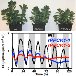
PPCK mutants (blue and red lines) are unable to regulate CO2 uptake with a circadian rhythm.

Question: We wanted to know if PPCK would be a necessary component of engineering other plants to use CAM photosynthesis. We tested this by switching the gene off in the CAM plant from Madagascar Kalanchoë fedtschenkoi (Lavender Scallops).

Findings: We found that, for CAM to work properly, the cells must switch on PPCK each night. When we prevented Kalanchoë from making PPCK at night, the plants could only capture a third of the CO2 captured by the normal plants. In addition, we discovered that the plants that were unable to make PPCK each night had alterations in their internal cellular timekeeping mechanism, the circadian clock. In CAM plants, the circadian clock optimizes CO2 fixation and PPCK is one of the key ways that the cellular clock communicates time signals to control the CAM process. What was surprising was that switching off PPCK led to changes in the circadian clock itself. We are now working to understand whether (and how) changes in cellular metabolism resulting from the drop in dark CO2 capture were communicated to the internal clock within the leaf cells where CAM occurs.

Next steps: Scientists aim to tweak crop plants to use the CAM system to produce new crops that are better suited to growth on drought-prone lands. Our work demonstrates that ongoing efforts to engineer CAM photosynthesis into other plants will need to introduce PPCK to protect PEPC from inhibition. With PPCK working the night shift, CAM works three times better, which should help crop scientists to make the most of this amazingly water-wise form of photosynthesis.

Susanna F. Boxall, Louisa V. Dever, Jana Knerova, Peter D. Gould and James Hartwell. (2017). Phosphorylation of Phosphoenolpyruvate Carboxylase is Essential for Maximal and Sustained Dark CO2 Fixation and Core Circadian Clock Operation in the Obligate Crassulacean Acid Metabolism Species Kalanchoë fedtschenkoihttps://doi.org/10.1105/tpc.17.00301

Keeping Walls on Track

Schneider et al. explore how secondary cell walls are made. The Plant Cell 2017.  https://doi.org/10.1105/tpc.17.00309

Background: Plant cells are surrounded by robust cell walls that function as dynamic extracellular skeletons and protect plants against their environment. The cell walls make up the bulk of biomass and are utilized in a range of industrial applications, including feed, food, fuel and construction material. Understanding how plants produce cell walls is therefore of great importance. Water-transporting xylem cells produce walls that are rich in cellulose (a polysaccharide) and lignin (composed of polyphenols). Strikingly, the walls of xylem cells are produced in intricate patterns, for example bands or spirals, that allow xylem cells to withstand internal negative pressure necessary to transport water. These patterns are thought to be directed by the intracellular cytoskeleton, in particular microtubules, that guide cellulose synthesis.

Connections between microtubule cytoskeleton and cellulose synthesizing proteins are needed for proper initiation of xylem vessel cell wall patterns. Panels show single cells in dark-grown seedling stems that express a fluorescently-tagged cellulose synthase (CESA; turquoise) that is active in xylem vessel formation and microtubule subunits (magenta).  In the early phases the CESA trajectories do not align well with the microtubules (see dotted lines and arrows), but the tracks become aligned with the microtubules over time (the fluorescence signals coincide in late stage). We conclude that the link between the CESAs and microtubules is needed only during the early stages of vessel formation and that another molecular process re-aligns the CESAs and microtubules during xylem differentiation.

Question: The components of the xylem walls are well known, but how the patterning of these walls is achieved is not well understood. We therefore set out to investigate how these patterns are established and sustained during xylem development.

Findings: We used several complementary systems to address these questions, including Arabidopsis and rice plants. One of the key experimental set-ups was the use of a xylem-inducible seedling system where we could directly image the process of xylem wall patterning using confocal microscopy. To do this we introgressed fluorescently-tagged proteins, with a focus on cellulose synthesizing proteins and microtubule subunits, into the inducible system and imaged these during xylem development. We also studied how xylem development and wall patterning are affected by mutations that disturb the connection between the microtubules and the cellulose synthesizing proteins. We found that microtubules are needed to establish wall patterns during xylem development, but that once the patterns are established they can be maintained without much contribution of the microtubules.

Next steps: While our research contributes important new information about how cell wall patterns are controlled, it is not clear what is driving the microtubules to change their distribution from a relatively evenly distributed array before xylem onset to a banded or spiralling pattern once the development is induced. In addition, another type of xylem cell has reticulated wall patterns and so it will be very interesting to identify similarities and differences in how these two patterns come about.

Rene Schneider, Lu Tang, Edwin R Lampugnani, Sarah Barkwill, Rahul Lathe, Yi Zhang, Heather E. McFarlane, Edouard Pesquet, Totte Niittyla, Shawn D. Mansfield, Yihua Zhou, Staffan Persson. 2017. CSI1/POM2 steers xylem vessel wall patterning. Plant Cell https://doi.org/10.1105/tpc.17.00309

To Grow or to Defend: That is the Question for Plant Central Metabolism

Fusari et al. perform GWAS to explore primary plant metabolism https://doi.org/10.1105/tpc.17.00232

By Corina M. Fusari and Rik Kooke

Background: Primary metabolites such as sugars, organic acids, and amino acids are essential chemical compounds that drive plant growth and development by providing the carbon and energy needed for cell division, expansion, and maintenance and for the production of stress and defense compounds. Primary metabolites form a strongly interconnected network, with individual metabolite levels determined by enzyme activities. Enzymes control the output of this network and ultimately, growth. Most plant characteristics, including metabolism, vary among different plant genotypes of the same species. Genetic mapping can be used to identify genes controlling variable features using a mapping population, typically obtained either by crossing two individuals (biparental mapping) or by using a natural population of individuals that shows variation in the targeted feature (genome-wide association studies, GWAS). Individuals from the population are then analyzed for the trait of interest and genotyped with molecular markers. Biparental mapping has been used to identify genes controlling metabolite levels, and occasionally enzymes. Enzyme abundance can be regulated by changes in genes encoding individual enzymes (in cis) or in other genes (in trans). While all metabolic components are highly coordinated, it remains unclear what generates the strongly connected metabolite networks observed in mapping populations.

Question: We performed GWAS in Arabidopsis thaliana to identify new genes involved in regulating plant primary metabolism and to search for regulatory hubs that generate coordinated changes in many metabolic traits.

Findings: We identified polymorphisms (DNA marker variations) in five genes (UGP1, VAC-INV, APL1, SIS, FUM1) responsible for variation in the activity of enzymes involved in sucrose synthesis and degradation, starch synthesis, and organic acid synthesis. For UGP1, two different sequences (alleles) coexist, leading to individuals with high and low enzyme activity; both have been maintained throughout evolution. For VAC-INV, the differences in activity levels result from combined mutations in its promoter and coding region that have occurred at different times in evolution. In addition, ugp1, vac-inv, and apl1 mutants have altered seed phenotypes. This suggests that seed development is especially sensitive to minor lesions in metabolism that have little effect at other stages in the plant’s life. We identified genes involved in transport, protein interactions, and protein degradation that regulate specific metabolites or enzymes (trans-regulation). We also found 14 genes controlling various metabolic components at the same time (pleiotropic regulation). The strongest pleiotropic hub was found at ACCELERATED CELL DEATH6 (ACD6), affecting six enzyme activities, three metabolites, protein, and biomass. ACD6 was previously linked to plant defense, spontaneous cell death, and plant biomass. We now know that ACD6 strongly contributes to the natural genetic variation in central metabolism in Arabidopsis, revealing a compromise between metabolism and defense against biotic stress.

Fusari, C.M., Kooke, R., Lauxmann, M.A., Annunziata, M.G., Encke, B., Hoehne, M., Krohn, N., Becker, F.F.M., Schlereth, A., Sulpice, R., Stitt, M., and Keurentjes, J.J.B. (2017). Genome-wide Association Mapping Reveals that Specific and Pleiotropic Regulatory Mechanisms Fine-tune Central Metabolism and Growth in Arabidopsis. Plant Cell https://doi.org/10.1105/tpc.17.00232

How Meiotic Chromosomes Cluster into a “Bouquet”

Zhang et al. explore the behavior of chromosomes during meiosis. The Plant Cell (2017). https://doi.org/10.1105/tpc.17.00287

By Fanfan Zhang

Background: In meiosis, plants, animals, and fungi reduce their chromosome numbers by half to form gametes (sperm and eggs) that can fuse to form a cell with a complete set of genetic material. During one stage of meiosis, chromosomes huddle together and the chromosome ends (telomeres) cluster in a limited area of the inner nuclear membrane. This cluster of chromosomes looks like an attractively arranged bunch of flowers, so it is widely called the “chromosomal bouquet”. Chromosomal bouquet formation happens in nearly all plants, animals and fungi, and plays an important role in pairing of the chromosomes and therefore progression of meiosis.

Question: In recent years, work in yeast and mammals has identified some essential factors participating in bouquet formation, but they are not conserved across species. So far, the underlying molecular mechanism in plant species remains largely unknown. So, we wanted to know what leads to the aggregation of chromosomes during meiosis in plants.

Findings: Based on a genetic screen for sterile mutants in rice (Oryza sativa), we identified ZYGOTENE 1 (ZYGO1), a novel protein that regulates bouquet formation during early meiosis. The zygo1 mutants fail to form proper bouquets and the mutant rice therefore lack typical meiotic cells at this stage. Because the plants cannot form proper gametes, they are sterile. Staining of the proteins on chromosomes in meiosis showed that the polarized localization of a protein that promotes telomere movement is also altered in the zygo1 mutants. All of these results suggest that ZYGO1 plays an essential role during telomere bouquet formation. We also found that ZYGO1 affects chromosome behavior independently of double-strand break formation and recombination. In addition, the failure chromosome clustering in the zygo1 mutants further affects other aspects of chromosome behavior, including homolog pairing, synaptonemal complex assembly, and crossover formation. ZYGO1 encodes an F-box protein, containing an F-box domain with some conserved residues. Our results showed that ZYGO1 interacts with the rice SKP1-like protein 1 (OSK1) through its F-box domain, suggesting that ZYGO1 modulates bouquet formation as a component of cellular factors involved in breaking down proteins.

Next steps: This investigation lays an important foundation for uncovering the molecular mechanism underlying chromosome clustering during meiosis. Further research towards identification of the target(s) of ZYGO1 will shed light on its specific function.

Fanfan Zhang, Ding Tang, Yi Shen, Zhihui Xue, Wenqing Shi, Lijun Ren, Guijie Du, Yafei Li, and Zhukuan Cheng (2017) The F-box protein ZYGO1 mediates bouquet formation to promote homologous pairing, synapsis, and recombination in rice meiosis. tpc.00287.2017; https://doi.org/10.1105/tpc.17.00287

Space-Time Continuum of Gene Expression in Lateral Root Development

Walker et al. explore how the environment shapes root architecture. The Plant Cell 2017.  https://doi.org/10.1105/tpc.16.00961

By Liam Walker

Background: To acquire nutrients and anchor themselves, plant roots spread both vertically and horizontally in soil. Plants typically have a primary root that grows vertically and lateral roots that arise from the primary root and grow horizontally. The plant root consists of many different types of cells organized in concentric layers around the core of the root. For example, pericycle cells form a layer close to the centre of the root, whereas cortex cells are closer to the outside of the root. The emergence of new lateral roots, depend on the action of specific types of cells, orchestrated by cell-specific changes in gene expression over time.

Question: Our aim was to understand how changes in the environment that affect root architecture are regulated by gene expression changes in different cell types over time.

Findings: We used Arabidopsis plants that contained a gene encoding a green fluorescent protein expressed either only in the cortex or only in the pericycle root cell types. We treated seedlings with either the nutrient nitrogen or a bacterium called Sinorhizobium meliloti, both of which alter lateral root formation, and harvested roots at many time points over two days after treatment. Next, we used enzymes to digest cell walls and detach individual cells from one another, then isolated individual cells using a cell sorting machine able to detect cells expressing fluorescent protein, and then measured gene expression in the isolated cells. We found that thousands of genes changed their expression patterns during the early stages of building new lateral roots, and the genes that showed changes were very different between the two cell types studied. Despite these differences, we found many similarities between the functions that these distinct sets of genes enacted between these different types of cells and between the treatments. This means that although the activity of the protein produced from a gene is important, both the location and timing of gene expression also contribute to its function.

Next steps: This work shows that studying how genes behave across time and space (in different cell layers) is crucial for fully understanding how plants function. It also provides other researchers studying lateral root development and plant environmental responses with an important resource.

Walker, L., Boddington, C., Jenkins, D.J., Wang, Y., Grønlund, J.T., Hulsmans, J., Kumar, S., Patel, D., Moore, J., Carter, A., Samavedam, S., Bonomo, G., Hersh, D.S., Coruzzi, G.M., Burroughs, N.J., and Gifford M.L. (2017). Changes in Gene Expression in Space and Time Orchestrate Environmentally-Mediated Shaping of Root Architecture. Plant Cell https://doi.org/10.1105/tpc.16.00961

A Plant Protein That Foils Aphid Feeding

Kloth et al. probe aphid feeding behavior. The Plant Cell 2017  doi: 10.1105/tpc.16.00424

By Karen Kloth

Background: Aphids are phloem-feeding insects. They penetrate plants with a piercing-sucking mouth. Once they reach a tube where the plant transports its sugar-rich phloem sap, they can take up sap for hours or even days. Unless the plant has a smart way to defend itself.

Question: To explore natural plant defense against aphids, we studied aphid feeding behavior on 350 varieties of the model plant Arabidopsis thaliana, originating from different locations in the northern hemisphere. To track aphid behavior on all these plants, we built a video-tracking platform. In this platform, aphids are released on leaf discs and recorded on video. Using special software, aphid feeding behavior was monitored on the 350 Arabidopsis varieties.

Findings: The aphids probed less on some plants than on others. This behavior was associated with natural mutations in one specific plant gene, coding for a heat shock-like protein with unknown function. To test if this protein really affects aphids, we screened aphid feeding behavior on mutant plants without the protein. On these plants, aphids ingested phloem sap longer and faster, and produced more offspring. The effects were particularly large at high temperature, 26 degrees Celsius, when plants produced more of the protein. These results confirmed that this protein increases plant resistance to aphids during heat stress. The protein also had another advantage. Plants with the protein were able to produce more seeds during heat stress than plants that lacked it.

Next, we studied where the protein is located. We transgenically expressed a fluorescent version of the protein in plants, and found that it is located in the tubes where phloem sap is transported. It coats the inside of the phloem tubes and forms spherical bodies around organelles known as mitochondria. In cell cultures, the location of the protein overlapped with another organelle, the endoplasmic reticulum. We think that the protein occludes the narrow food canal of the aphid and thereby obstructs phloem sap uptake. In addition, it might increase sap transport to the flower and seeds during heat stress, but that needs to be verified.

Next steps: Natural plant resistance to aphids and better tolerance to heat stress are of interest for plant breeding companies. Breeding crops with effective resistance proteins can help to reduce insecticide application and yield losses due to hot conditions. In the long term, this research could help farmers produce more sustainable fruits and vegetables.

Kloth, K.J., Busscher, J., Wiegers, G., Kruijer, W., Buijs, G., Meyer, R.C., Albrectsen, B.R., Bouwmeester, H.J., Dicke, M., and Jongsma, M.A. (2017). SIEVE ELEMENT-LINING CHAPERONE 1 restricts aphid feeding on Arabidopsis during heat stress. Plant Cell. https://doi.org/10.1105/tpc.16.00424

Videos of aphid probing behavior:


Flowering Versus Runnering: A Very Important Decision in Strawberry

Tenreira et al. find a gene responsible for the differentiation of the stolon in strawberry. The Plant Cell 2017. https://doi.org/10.1105/tpc.16.00949

Asexual reproduction produces offspring that are genetically identical to the parents. This process takes many forms in flowering plants, including the production of tubers, rhizomes, corms, bulbs, and stolons. In strawberry, stolons , (also known as runners) are elongated stems that produce daughter plants that are used to propagate commercial varieties. The axillary meristem (embryonic tissue in the leaf axil) gives rise to stolons, but it can also produce inflorescence-bearing shoots, leading to flowering and fruit production. Until recently, little was known about the mechanism underlying the flowering/runnering decision in strawberry.

Tenreira et al. (2017) used a natural runnerless (r) mutant to identify the RUNNERING gene (responsible for stolon production) in woodland strawberry. Using a classical genetics approach combined with mapping via next-generation sequencing, the r mutation was identified as a deletion in the active site of the Gibberellin 20-oxidase4 (GA20ox4) gene, which is expressed primarily in the axillary meristem. GA20ox is a key biosynthetic enzyme that catalyzes a rate-limiting step in the synthesis of bioactive gibberellins (GAs). GAs are phytohormones that play a central role in many developmental processes. In strawberry, as in other perennial species, GAs inhibit flowering and promote vegetative growth.

Tenreira et al. demonstrated that GA20ox4 is strictly linked to stolon formation in the axillary meristem. When this protein is mutated, its enzymatic activity is totally lost. Depending on the allelic state of GA20ox4, the axillary meristem produces either a stolon (active allele) or an inflorescence-bearing shoot as the default developmental program (inactive allele). Therefore, GA20-oxidase plays a pivotal role in the decision of the meristem to produce either stolons or inflorescence-bearing shoots, and thus in the trade-off between flowering and runnering in diploid strawberry.

It will be interesting to explore regulatory genes and targets of GA in the axillary meristems of strawberry. A future challenge is to transfer this knowledge to cultivated strawberry in order to modulate the balance between runnering and flowering in this important crop.

Tenreira, T., Pimenta Lange, M.J., Lange, T., Bres, C., Labadie, M., Monfort, A., Hernould, M., Rothan, C., and Denoyes, B. (2017). A specific gibberellin 20-oxidase dictates the flowering-runnering decision in diploid strawberry. Plant Cell 29: https://doi.org/10.1105/tpc.16.00949

Light Helps Plants Cope with Phosphate Starvation

Liu et al. focus on transcriptional regulation of PHR1 expression. The Plant Cell 2017. https://doi.org/10.1105/tpc.17.00268

Phosphorus (P) is an essential micronutrient for plant growth, development, and metabolism. Phosphate (Pi), the major form of P used by plants, is highly immobile in most soils, making it one of the most limiting nutrients for crop productivity. The overuse of P fertilizers has been rapidly depleting P resources and causing severe damage to the ecosystem. It is crucial to breed crops that can take up and use P more efficiently, but such efforts have so far met only limited success since we know little about the molecular mechanisms regulating Pi signaling and responses in plants.

To cope with Pi deficiency, plants have evolved many ways to remobilize and redistribute internal Pi and to better acquire Pi from the soil. Pi starvation responses include remodeling of 
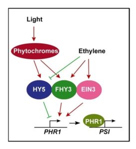
root system architecture (stopping primary root growth and increasing root hair and lateral root formation), reducing photosynthesis, enhancing Pi transporter activity, and accumulating starch and the pigment anthocyanin. How these activities are orchestrated is not yet well known. Arabidopsis PHOSPHATE STARVATION RESPONSE1 (PHR1) encodes a conserved transcription factor that helps program Pi starvation-induced gene expression and the resulting downstream Pi starvation responses. However, PHR1 expression is only weakly responsive to Pi deprivation stress. In this work, Liu et al. (2017) show that PHR1 expression is induced by light in a manner dependent on light receptors known as phytochromes.

FHY3, FAR1, and HY5, three positive regulators of the phytochrome light-signaling pathway, directly bind to the PHR1 gene promoter. Strikingly, FHY3 and FAR1 activate, while HY5 represses, PHR1 expression. In addition, EIN3, a master regulator of the plant hormone ethylene, also directly binds to the PHR1 promoter and activates its expression. FHY3 directly interacts with EIN3, and HY5 can suppress the activation of PHR1 by FHY3 and EIN3. Finally, both light and ethylene promote FHY3 protein accumulation, and ethylene blocks the light-promoted stabilization of HY5. Therefore, it appears that light and ethylene coordinately regulate PHR1 expression and the phosphate starvation response through signaling convergence at the PHR1 promoter. These findings provide new insights into the molecular mechanism governing how light and ethylene together regulate Pi signaling and the phosphate starvation response. Such findings may ultimately help breeders design crop varieties with an improved ability to acquire and utilize Pi.

Liu, Y., Xie, Y.-R., Wang, H., Ma, X.-J., Yao, W.-J., Wang, H.-Y. (2017). Light and Ethylene Coordinately Regulate the Phosphate Starvation Response through Transcriptional Regulation of PHOSPHATE STARVATION RESPONSE1. Plant Cell DOI: https://doi.org/10.1105/tpc.17.00268.


How Does Histone Phosphorylation Affect Flowering Time?

Su et al. look at chromatin modifications that affect flowering. The Plant Cell 2017. https://doi.org/10.1105/tpc.17.00266

Plants, unlike animals, begin their lives as seeds that – in flowering plants – develop from flowers. This depends upon proper regulation of flowering time, to ensure pollination, fertilization, and development of the seed at the right time. In the long-day plant Arabidopsis thaliana, GIGANTEA (GI) protein activates CONSTANS (CO), which in turn upregulates FLOWERING LOCUS T (FT) to promote flowering under a long-day photoperiod. Thus regulation of GI is crucial to the process. CIRCADIAN CLOCK ASSOCIATED1 (CCA1) binds to GI and blocks its activation, but the activators of GI remain unclear.

In eukaryotic cells, covalent modifications in chromatin, such as histone phosphorylation and acetylation, participate in development by activating or blocking transcription of genes. Plants might have specific histone modification sites if chromatin truly contains developmental information. A primary question is how plant-specific histone modifications and other regulators work together to promote proper flowering time in Arabidopsis.

Su et al. found that the protein MUT9P-LIKE-KINASE (MLK4) promotes proper flowering time under a long-day photoperiod. MLK4 phosphorylates the histone H2A at serine 95, a site that exists in plants, but not in yeast, Drosophila, mouse, or humans. CCA1 interacts with MLK4, allowing it to to bind GI. Moreover, in vivo, MLK4 interacts with YAF9a, a subunit of a large protein complex that incorporate the histone variant H2A.Z into chromatin. The Arabidopsis mlk4 mutant exhibits a reduction in the levels of serine 95 phosphorylation of histone H2A, H2A.Z enrichment, and GI transcript, which in turn delays flowering time.

Curiously, MLK4 has no function in regulating flowering time under a short-day photoperiod. The authors hypothesized that MLK4 might have different mRNA levels or activities under long-day versus short-day photoperiods. Future studies will focus on understanding how this enzyme is regulated by photoperiod, because only plants that flower at the right time can generate seeds.

Su, Y.H., Wang, S.L., Zhang, F., Liu, Y.N., Huang, T.T., and Ding, Y. (2017) Phosphorylation of Histone H2A at Serine 95: a Plant-specific Mark Involved in Flowering Time Regulation and H2A.Z Deposition. Published August 2017. https://doi.org/10.1105/tpc.17.00266.