Sending out a salt-related SOS: transcriptional regulation of salt stress responses

Lu et al. explore the roles of a Mediator complex subunit and a WRKY transcription factor in regulating the salt-responsive gene SOS1.

https://doi.org/10.1093/plcell/koad105

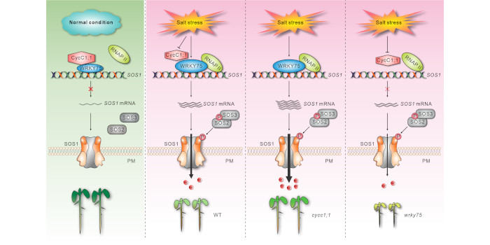
Background: Soil salinization is a major environmental hazard that severely affects plant growth and development. Plants have evolved sophisticated mechanisms that help them withstand elevated soil salinity, and Salt Overly Sensitive1 (SOS1) plays a crucial role in plant salt stress tolerance by facilitating the extrusion of excess Na+ from the cells. Although previous reports have demonstrated the important role of post-translational regulation of SOS1 in plant salt stress tolerance, how SOS1 transcription is dynamically modulated in response to different salinity conditions remains unclear.

Question: What are the molecular mechanisms by which plants regulate SOS1 expression at the transcriptional level in response to salinity stress?

Findings: Disruption of the CycC1;1 subunit of the plant Mediator complex promotes salt-induced SOS1 expression and salt tolerance in Arabidopsis because CycC1;1 interferes with RNA polymerase II recruitment by occupying the SOS1 promoter. SOS1 mutation in the cycc1;1 mutant completely compromised its enhanced salt tolerance. Moreover, CycC1;1 can physically interact with the transcription factor WRKY75, which can directly bind to the SOS1 promoter and activate its expression. In contrast to the cycc1;1 mutant, the wrky75 mutant has attenuated SOS1 expression and salt tolerance, whereas overexpression of SOS1 can rescue the salt sensitivity of the mutant. Intriguingly, CycC1;1 inhibits WRKY75 transcriptional activation activity for SOS1 through their interaction; thus, increased SOS1 expression and salt tolerance in the cycc1;1 mutant was abolished by the WRKY75 mutation. In addition, CycC1;1 expression is repressed and WRKY75 expression is stimulated in response to high salinity.

Next steps: In a future study, we will explore whether other components of the Mediator complex are coordinated with CycC1;1 to precisely control SOS1 transcription, and investigate how salinity affects CycC1;1 and WRKY75 expression in the plant’s response to different salinity co

Reference:

Kai-Kai Lu, Ru-Feng Song, Jia-Xing Guo, Yu Zhang, Jia-Xin Zuo, Hui-Hui Chen, Cai-Yi Liao, Xiao-Yu Hu, Feng Ren, Ying-Tang Lu and Wen-Cheng Liu (2023) CycC1;1-WRKY75 complex-mediated transcriptional regulation of SOS1 controls salt stress tolerance in Arabidopsis. https://doi.org/10.1093/plcell/koad105

Cryptophyte PSI–LHCI supercomplexes: understanding the structural diversity and evolution of PSI–LHCI in the red lineage

Zhao, Wang, Li, et al. explore how the photosynthetic apparatus evolved in the cryptophyte Chroomonas placoidea, which is related to red algae.

Long-Sheng Zhao1 and Lu-Ning Liu2.

1State Key Laboratory of Microbial Technology, Shandong University, Qingdao, China.

2Institute of Systems, Molecular and Integrative Biology, University of Liverpool, Liverpool L69 7ZB, United Kingdom.

 Background: Photosynthesis converts solar energy into biologically useful energy and generates oxygen, sustaining almost all life forms on Earth. Photosystem I (PSI) is a large pigment-protein supercomplex that plays a key role in photosynthesis. High-resolution structural analysis of the PSI and its antenna complexes is crucial for elucidating the mechanism of light capture and energy transfer in photoautotrophs. So far, the PSI structures of cyanobacteria, red algae, green algae, diatom, moss, and land plants have been resolved, providing insights into the energy conversion mechanisms and important clues for the evolutionary diversity of PSI–LHCI structures.

Question: Cryptophytes are an ancient group that originated from red algae during secondary endosymbiosis. They possess unique evolutionary traits and play a crucial role in ecology. Despite their importance in ecological function and evolution, the high-resolution structure of cryptophyte PSI remains elusive.

Findings: The PSI–light-harvesting antenna (LHCI) supercomplex was isolated from a cryptophyte Chroomonas placoidea and its structure was determined by cryo-EM. Cryptophyte PSI is composed of 14 core subunits, 14 LHCIs surrounding the PSI core and a pigment-binding polypeptide not present in any other reported PSI–LHCI complexes. The structure coordinates a total of 373 pigments which facilitate efficient capturing of light and energy transfer from LHCIs to the PSI core. Cryptophyte PSI–LHCI shares common structural features with both red algal and diatom counterparts, while also displaying unique protein organization, pigment association, and energy transfer pathways. By uncovering these structural variations, our study sheds light on the unique features of cryptophyte PSI–LHCI as an intermediate state during the evolution of red lineage PSI–LHCI.

Next steps: Our cryo-EM structure reveals an intense pigment network within cryptophyte PSI–LHCI. Comprehensive and accurate analysis of energy transfer pathways in cryptophyte PSI–LHCI relative to the PSI–LHCI from other photoautotrophs needs to be performed experimentally and theoretically. Moreover, uncovering the PSI–LHCI structures of various species is critical to understanding the structural diversity of PSI–LHCI during evolution and environmental adaptation.

Reference:

Long-Sheng Zhao, Peng Wang, Kang Li, Quan-Bao Zhang, Fei-Yu He, Chun-Yang Li, Hai-Nan Su, Xiu-Lan Chen, Lu-Ning Liu, Yu-Zhong Zhang (2023) Structural basis and evolution of the photosystem I–light-harvesting supercomplex of cryptophyte algae. https://doi.org/10.1093/plcell/koad087

The importance of being in the right place to respond to light cues

Lopez Vazquez et al explore the importance of protein sub-cellular localization by determining how a protein modulating light responses gets to the right place within the cell.

Christian Fankhauser, Centre for Integrative Genomics, Faculty of Biology and Medicine, Génopode Building, University of Lausanne, CH–1015 Lausanne, Switzerland

Background: Plants can orient their leaves towards the light. This is known as phototropism and fascinated scientists since Charles Darwin. Phototropism is initiated by a blue light photoreceptor called phototropin. Light activates phototropin’s protein kinase activity (an enzyme that phosphorylates proteins). This is followed by a series of poorly understood events, which lead to the redistribution of the growth hormone auxin in the stem resulting in growth towards the light. PHYTOCHROME KINASE SUBSTRATE 4 (PKS4) is phosphorylated by phototropin and is believed to act between phototropin light activation and auxin redistribution. The mechanism of PKS4 action is unknown because the protein does not contain any domains of known biochemical activity. PKS4 associates with the plasma-membrane, where it was postulated to act.

Question: We wanted to understand how PKS4 associates with the plasma membrane and determine whether this subcellular localization is important for its biological activity.

Findings: Functionally important parts of proteins are typically conserved over evolutionary timescales. We therefore identified genes coding for PKS proteins and found that they are present in seed plants and comprise 6 conserved sequence motifs. We showed that one of these motifs is important for efficient PKS association with the plasma membrane in Arabidopsis. Cysteine residues of this motif are modified with lipids, which presumably contribute to plasma membrane association. Moreover, in PKS4 these residues are important for its function in phototropin signaling in Arabidopsis.

Next steps: Our study identified a PKS4 sequence motif that is important for biological activity and subcellular localization. However, we still don’t know what the rest of the protein does and how it may be important in linking phototropin activation and auxin redistribution. We hope that studying other conserved motives identified here will allow us to answer this question.

Reference:

Ana Lopez Vazquez, Laure Allenbach Petrolati, Martina Legris, Christophe Dessimoz, Edwin R. Lampugnani, Natasha Glover, and Christian Fankhauser (2023) Protein S-acylation controls the subcellular localization and biological activity of PHYTOCHROME KINASE SUBSTRATE. https://doi.org/10.1093/plcell/koad096

Interdependence of subunits of the m6A methyltransferase complex

Lisha Shen uncovers unique functional interdependence at the post-translational level among individual components of the Arabidopsis m6A methyltransferase complex. 

By Lisha Shen, Temasek Life Sciences Laboratory and Department of Biological Sciences, National University of Singapore, Singapore

Background: N6-methyladenosine (m6A), methylation at the nitrogen-6 position of adenosine, is an abundant internal modification of messenger RNA (mRNA) in plants, animals, and fungi. Adding m6A to mRNAs requires an evolutionarily conserved multicomponent complex called the m6A methyltransferase complex. In the model plant Arabidopsis thaliana, this complex contains two core methyltransferases, mRNA adenosine methylase (MTA) and MTB, and several accessory subunits including FKBP12 INTERACTING PROTEIN 37KD (FIP37), VIRILIZER (VIR) and HAKAI. Among these components, MTA, MTB, FIP37, and VIR are essential for m6A methylation.

Question: Although several components have been identified in the m6A methyltransferase complex, how these components influence each other to fulfill the function of the m6A methyltransferase complex as a whole remains largely unknown.  

Findings: The individual components of the m6A methyltransferase complex mostly affect each other at the protein level rather than at the gene expression level. First, MTA and MTB affect each other’s protein abundance. Second, FIP37 and VIR stabilize MTA and MTB, thus maintaining m6A methyltransferase complex function. Third, VIR affects the protein accumulation of FIP37 and HAKAI. In contrast, HAKAI has little effect on the protein abundance of MTA, MTB and FIP37. These results suggest that modulation of protein levels of individual subunits at the post-translational level is essential for the proper function of the m6A methyltransferase complex in depositing m6A modifications in plants.

Next steps: In future studies, I will further investigate whether protein homeostasis among various subunits of the m6A methyltransferase complex is dynamically altered and regulated in different developmental contexts and in response to different environmental conditions.

Reference:

Lisha Shen. (2023). Functional interdependence of subunits of the m6A methyltransferase complex in Arabidopsis. https://doi.org/10.1093/plcell/koad070

Clathrin-mediated endocytosis of RxLR effectors into plant cells

Wang et al. discover that Phytophthora infestans RxLR effectors enter host plant cells via clathrin-mediated endocytosis.

By Haixia Wang1, Petra C Boevink2, Paul RJ Birch1,2

1Division of Plant Science, School of Life Sciences, University of Dundee, @James Hutton Institute, Errol Road, Invergowrie, Dundee DD2 5DA, UK.

2Cell and Molecular Sciences, James Hutton Institute, Errol Road, Invergowrie, Dundee DD2 5DA, UK.

Background: Disease causing oomycete pathogens, such as Phytophthora infestans, the cause of potato late blight, represent a threat to global food security. To establish disease P. infestans delivers virulence proteins called ‘RxLR’ effectors into living plant cells. RxLR effectors target host proteins and processes and manipulate them to suppress immunity.

Question: A key question addressed in our work is: are RxLR effectors taken up into plant host cells by clathrin-mediated endocytosis?

Findings: We explored whether clathrin-mediated endocytosis (CME) is involved in RxLR effector uptake in three ways. Firstly, we silenced key host components, clathrin itself and a RAB protein called Ara6, and found that both P. infestans infection and RxLR effector delivery into host cells were reduced. Secondly, we used labelled clathrin and Ara6 proteins to immunopurify endosomal vesicles from plant cells during infection and found that a tagged RxLR effector was co-purified. Thirdly, we again captured endosomal vesicles during infection and performed a proteomics study, demonstrating that RxLR effectors, but not ‘apoplastic’ effectors that function outside of plant cells, were associated with the purified endosomes.

Next steps: Uptake of RxLR effectors into plant cells by CME implies that, although inside the host cells, the effectors are nevertheless compartmentalised in membrane vesicles. How do they escape the confines of the endomembrane system to reach their host target proteins in different subcellular locations?

Reference:

Haixia Wang, Shumei Wang, Wei Wang, Lin Xu, Lydia RJ Welsh, Marek Gierlinski, Stephen C Whisson, Piers A Hemsley, Petra C Boevink, Paul RJ Birch. (2023). Uptake of oomycete RXLR effectors into host cells by clathrin-mediated endocytosis. https://doi.org/10.1093/plcell/koad069

Review: Strigolactones integrate metabolic and nutritional signals

Strigolactones are a class of hormones first identified in the 1960s as components of root exudates that promote germination of parasitic Striga seeds, and later as a promoter of associations with arbuscular mycorrhizal fungi. Strigolactones also have endogenous roles within plants, for example as regulators of shoot branching. More recently, they have been shown to be involved in metabolic and nutrient sensing and signaling. These latter roles are summarized in a new review by Barbier et al. Several studies have demonstrated that the synthesis of strigolactones is stimulated by nutrient deficiency. Interestingly, changes in sugar metabolites affect strigolactone signaling but not apparently its synthesis. The molecular interactions through which these effects are mediated are also discussed. Finally, the authors discuss the many ways through which strigolactones help plants adapt to unfavorable environmental conditions. (Summary by Mary Williams @PlantTeaching) Nature Plants  10.1038/s41477-023-01453-6

Evolution of cytosolic and organellar invertases empowered the colonization and thriving of land plants

Invertases catalyse the conversion of sucrose into glucose and fructose. In plants, invertases fall into two clades – the mitochondrially/plastid localized α clade and the cytoplasmically localized β clade. However, we do not fully understand how these clades evolved. Here Wan et al. identified 665 invertases from 77 species across the plant kingdom. Phylogenetic analysis showed that invertases first appeared in charophyte algae due to endosymbiotic gene transfer from a cyanobacterium. The α1 clade, a plastid localised subgroup of the α clade, are the most basal invertases. Examination of N-terminal sequences revealed that α1 invertases are approximately 100 bp longer than β clade invertases. Combining this with phylogenetic analysis revealed that the β clade arose from duplication and subsequent loss of the transit peptide of the α1 clade. This happened before land plants diverged from charophyte algae. The α1 clade underwent an additional duplication event as vascular plants emerged, which led to the generation of the mitochondrially localised α2 clade. Collectively these results shed light on invertase evolution, and the authors suggest this was important for allowing plants to colonize land. (Summary by Rose McNelly @Rose_McN)  Plant Physiol. 10.1093/plphys/kiad401

CPK32 regulates cellulose biosynthesis through post-translational modification of cellulose synthase

Cellulose in one of the most abundant polymers on the planet and is synthesized by plasma membrane-bound cellulose synthases (CESAs). Phosphorylation plays a role in CESA regulation, however the kinases which catalyse the phosphorylation are not well described. Here Xin et al. identified calcium-dependent protein kinase 32 (CPK32) as an interactor of CESA3 in a yeast two hybrid screen. In vitro kinase assays revealed that CPK32 phosphorylates the catalytic domain of CESA3. To investigate how CPK32 phosphorylation affects CESA activity, they mutated a lysine residue in the CPK32 ATP-binding site (CPK32ΔCK96M). This mutation prevented kinase activity but not the interaction with CESAs. When CPK32ΔCK96M was transformed into Arabidopsis thaliana plants expressing YFP-tagged CESAs, CESA-YFP movement decreased by 28%. There was also a decrease in CESA-YFP stability. Protein degradation experiments showed a 30% reduction in the amount of CESA-YFP when CPK32ΔCK96M was expressed. This suggests a novel role of phosphorylation in controlling CESA stability during cellulose biosynthesis. (Summary by Rose McNelly @Rose_McN) New Phytol. 10.1111/nph.19106

Gravity sensing involves LZY translocation from statoliths to the plasma membrane

In plants, gravity sensing is orchestrated by specialized cells called statocytes that contain starch-filled plastids (amyloplasts) called statoliths, whose positions are influenced by the direction of gravity. However, the molecular mechanisms of gravity sensing and signaling are poorly understood. In Arabidopsis, LAZY1-LIKE (LZY) family genes have been shown to be required for auxin redistribution in response to gravitropic stimulation. Nishimura et al. investigated the role of LZY3 polarization in gravity sensing and gravitropism.  The LZY3 protein, despite lacking a detectable transmembrane domain, appears to have basic hydrophobic clusters that can help with membrane association. The authors introduced glutamine substitutions into two sites to decrease the basic hydrophobic cluster. Altering the basic hydrophobic cluster at site A led to an accumulation of LZY3 in the basal region of the plasma membrane (PM), rescuing the primary root angle in the lzy2;3;4 triple mutant and demonstrating a role for membrane association in LZY3 function. The authors also showed that establishment of LZY polarity on the PM occurs by translocation of LZY from amyloplasts to the PM. All these together show the role of LZY as a signal molecule responsible for conveying the positional information of statoliths to the PM, which directly links gravity sensing to downstream signaling. (Summary by Andrea Gómez-Felipe @andreagomezfe) Science 10.1126/science.adh9978