Imaging plant germline differentiation within Arabidopsis flowers by light sheet microscopy (eLIFE)
 Advances in microscopy have greatly informed our understanding of fundamental plant processes, but the germline cells in flowers have been hard to image as they are tiny and embedded within other tissues. Valuchova et al. present a method using light sheet fluorescence microscopy that allows live cell imaging (for up to five days) of germline cells in Arabidopsis flowers. This allowed them to trace male meiosis within anthers, using a fluorescently-tagged histone H2A to image chromosomes, or they meiotic chromosome marker ASY1. Sepals were removed, and then flowers were embedded in low-melting point agarose in a capillary to hold them in place during imaging. Imaging female meiosis required further dissection to expose the ovules. This paper has some exciting time-lapse videos – see for example https://elifesciences.org/articles/52546#fig3video1 (Summary by Mary Williams) eLIFE 10.7554/eLife.52546
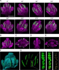