Structural and functional imaging of large and opaque plant specimen (J Exp Bot)
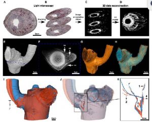 Even the simplest organisms are highly complex systems in which countless dynamic biochemical processes occur simultaneously. To fully understand the mechanical significance of such a complex molecular machine, the ability to accurately characterize dynamic processes at different scales is required. A variety of imaging methodologies are used to collect data for quantitative studies of plant growth and development. Multi-level data can be acquired, from macroscopic (m to cm) to molecular (nm), and from weeks to seconds. Static (at a given time; 3D) and dynamic (at different time points; quasi 4D) structural and functional imaging are a prerequisite for spatially resolving the form-structure-function relationships in plants. Each imaging technique has specific advantages and limitations as well as the appropriate fields of application. In this article, Hesse et al. described three different methods (LMTS, µ-CT and MRI) used in plant sciences, making a comparison of the imaging principles with a particular focus on their use in imaging large and opaque plant specimens. (Summarized by Francesca Resentini) J. Exp. Bot. 10.1093/jxb/erz186