
Review. Overcoming the scale barrier: expansion microscopy for nanoscale imaging in plants
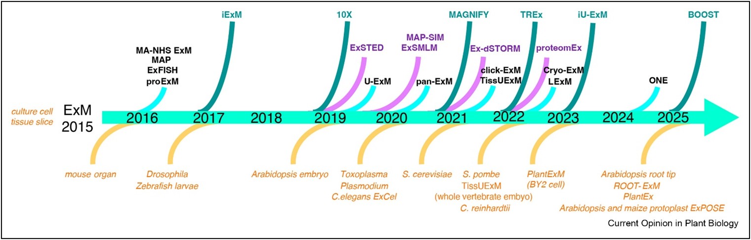 Many biological questions, from tissue-level patterning to subcellular organization, demand 3D imaging with spatial resolution beyond the limits of conventional light microscopy, or even super-resolution microscopy. Expansion microscopy (ExM) offers an elegant solution by physically enlarging biological specimens. By embedding samples in a swellable polymer gel, ExM typically expands biological structures by four- to twenty-fold, enabling nanoscale features to be resolved. In a recent review, Bayer and Grison summarized the development of ExM, outlining its core workflow: chemical crosslinking of biological components to a gel matrix, proteolytic digestion to soften the sample, and isotropic swelling of the gel to preserve structural relationships at an expanded scale. They also critically discuss current limitations and technical hurdles, with particular emphasis on plant science. Rigid cell walls, complex multicellular organization, low expansion factors, and uneven labeling remain major challenges. Consequently, ExM applications in plants have so far been restricted to Arabidopsis root tips, protoplasts, cultured cells, or young tissues lacking secondary wall modifications. Looking ahead, continued methodological innovation will be the key to unlock the full potential of ExM for visualizing plant structures across scales and developmental stages. (Summary by Ching Chan @ntnuchanlab @ntnuchanlab.bsky.social) Curr. Opin. Plant Biol. 10.1016/j.pbi.2025.102848