
An antibody-based probe enables visualization of active viral replication in living cells
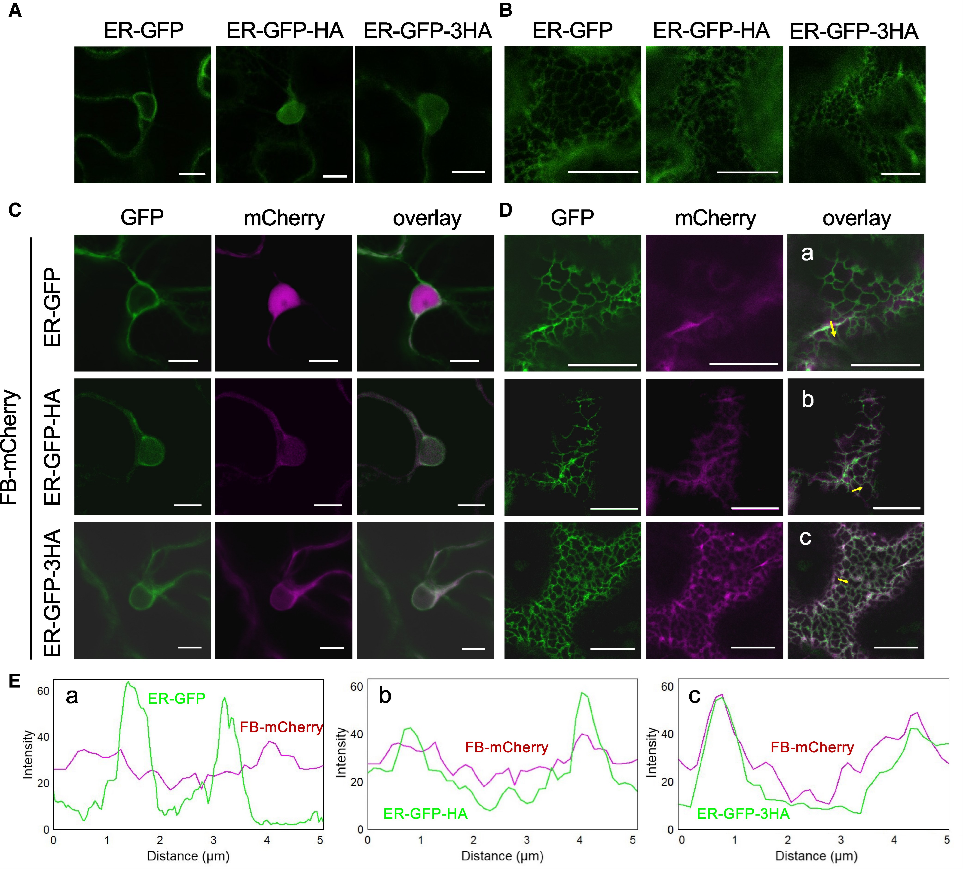 One of the main challenges in plant virology is detecting the active replicase enzyme of positive-strand RNA viruses in living cells, since adding fluorescent tags often prevents it from functioning. A recent study in Plant Physiology by Ishihara et al. has found a way around this problem. The researchers adapted the human influenza hemagglutinin (HA) frankenbody (FB), a genetically encoded single-chain antibody fragment that binds to a small HA epitope, for use in plant cells. They used a two-part, virus replication-inducible system so the FB-mCherry probe was expressed only in cells infected with Plantago asiatica mosaic virus (PlAMV). This approach kept probe levels under control and avoided background signals that could hide the specific labeling of an HA-tagged, fully functional viral replicase. Using live-cell imaging, they saw that the active PlAMV replicase forms small, distinct structures near the endoplasmic reticulum and plasmodesmata. These labeled granules were next to spots of double-stranded RNA, which is a sign of active viral replication, confirming they are real viral replication complexes. This FB-based system is the first flexible method for watching a working plant virus replicase in real time, and it could help researchers study how these complexes form and interact with the host cell. (Summary by Jahed Ahmed) Plant Physiol. 10.1093/plphys/kiaf240