A close-up view of the thylakoids (eLIFE)
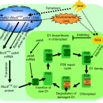
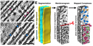 The thylakoid membranes inside the chloroplasts house the major protein complexes required for photosynthesis, including photosystems I and II (PSI/II), the b6f complex and ATP synthase. To optimize photosynthetic efficiency, the distribution and abundance of these complexes are dynamically regulated in response to varying light and growth conditions. However, to study how the spatial organization and composition of thylakoids contributes to photosynthetic productivity is challenging. Here, Wietrzynski et al. introduce “membranograms”, a cryo-EM-based method for mapping protein complexes onto segmented thylakoid membranes. This approach, here used on the green alga Chlamydomonas, gives an in situ, molecular-scale view of thylakoid architecture. Thylakoids fall into two distinct categories: appressed and non-appressed. Appressed membranes are stacked closely (~ 3 nanometer) together, whereas non-appressed ones are farther apart, and this structural distinction is a major determinant for which protein complexes are present. For example, the authors find that PSII is entirely confined to the appressed regions, whereas PSI and the ATP synthase occupies the non-appressed regions, with essentially no “mixing” at the borders. In contrast, the b6f complex, which is an important intermediary between PSII and PSI is more evenly distributed throughout the thylakoids. It will be interesting to see how this, and other properties of the thylakoid network discussed in the paper, changes when cells are challenged with different light conditions. (Summary by Frej Tulin @FrejTulin) eLIFE 10.7554/eLife.53740
[altmetric doi=”10.7554/eLife.53740″ details=”right” float=”right”]