
Unveiling vacuole biogenesis: Tubular networks are present in plant meristem cells
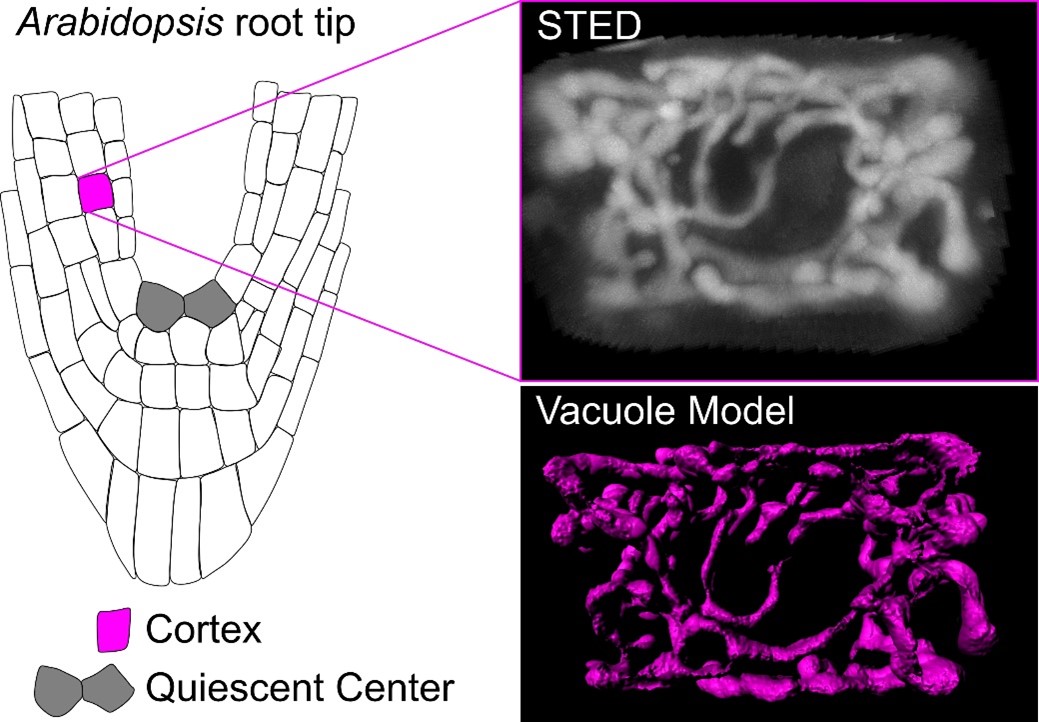 A recent paper by Scheuring and colleagues investigates vacuole biogenesis in meristematic cells of Arabidopsis thaliana, challenging earlier models of vacuole formation. Vacuoles are crucial organelles responsible for various cellular functions, yet their formation has remained puzzling for quite some time. Historically, models have proposed that vacuoles are formed either by contributions from the endoplasmic reticulum (ER) or through homotypic fusion of multivesicular bodies (MVBs). However, recent evidence suggests a more complex process involving multiple membrane sources. The authors use advanced imaging techniques, such as super-resolution stimulated emission depletion (STED) microscopy and fluorescence recovery after photobleaching (FRAP), to confirm the presence of a tubular vacuolar network in meristematic plant cells. The authors introduced a RUBY vacuole reporter line (using betalain fluorescence) to image deeper into tissues, where conventional dyes might not penetrate. This tubular network, observed in young cells near the quiescent center of the root, appears interconnected, which contradicts the theory that MVBs alone are sufficient to form vacuoles. Their findings suggest instead that vacuolar membranes originate from various cellular sources, including the ER. The study further introduces a customized FRAP assay, termed vacuole-connectivity FRAP (vaccFRAP), to analyze vacuolar dynamics. This assay beautifully highlights different connectivity levels within the vacuolar network, providing insights into how vacuoles function as interconnected compartments even in early developmental stages. This research advances our understanding of vacuole formation and its role in plant cell development. (Summary by Ann-Kathrin Rößling @AK_Roessling) Plant Cell 10.1093/plcell/koae243


