
Plant Science Research Weekly: October 31, 2025
Opinion: Genomic studies hint at what makes a tree a tree
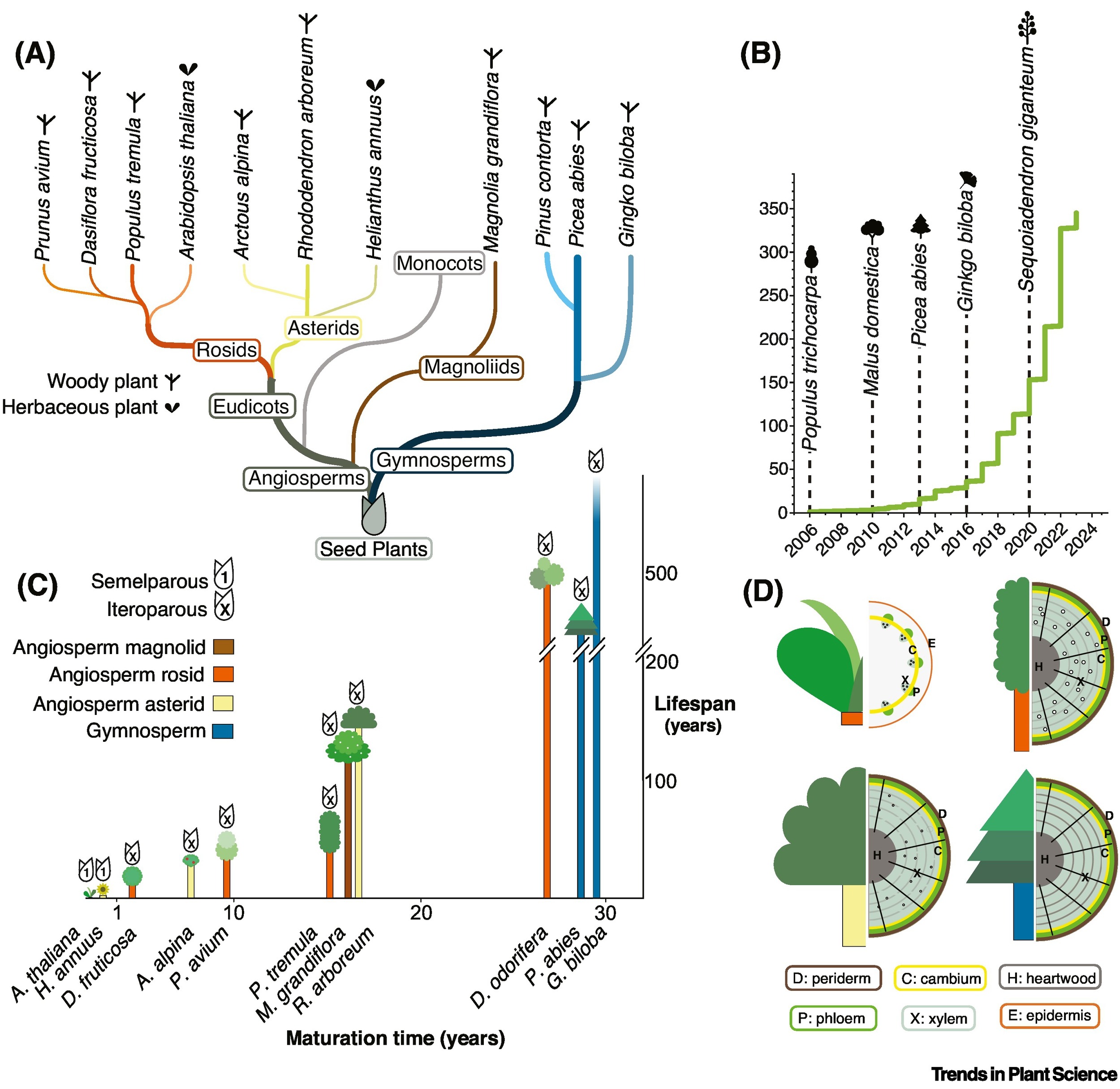 This is such an engaging and though-provoking review article. I’d love to ask a group of students to think about what makes a tree a tree, including such questions as: How do we define trees? Do woody plants share a single origin? How many angiosperms are woody? The answer to those questions and more can be found in this article by Birkeland et al., which draws on comparative studies of related herbaceous and woody species. Trees are defined by their woody secondary growth, which enables them to be longer-lived and bigger than herbaceous plants, usually accompanied by repeated cycles of reproduction. Although the potential to be woody is thought to be ancestral in angiosperms, it is clear that woodiness has been lost and regained repeatedly, suggesting that this trait is controlled by a small number of genes. Following extensive analysis of woody plant genomes and their relatives, the authors identified several genes that associated with the longer lifespans of trees include those that control the length of the juvenile phase, time of flowering, and stress resilience. The authors conclude with a set of outstanding questions, including “Why do angiosperms display such a wide variation of growth forms compared with gymnosperms, and is this variation linked to their evolutionary success?”. What a lot of fascinating things to think about! (Summary by Mary Williams @PlantTeaching.bsky.social). Trends Plant Sci 10.1016/j.tplants.2025.09.006
A new shade of photosynthesis: The missing chlorophyll f found in action
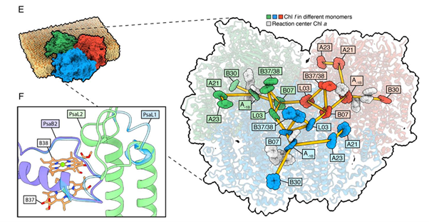 Photosynthetic organisms were long believed to only use visible light for energy capture, until the discovery of far-red photosynthesis challenged this view. Far-red photosynthesis is thought to be enabled by the presence of the pigment chlorophyll f. Although previous studies have identified chlorophyll f within Photosystem I of cyanobacteria, its precise photochemical function has remained unclear. Consoli et al. now provide a structural breakthrough by resolving two high-resolution (1.89 Å and 2.01 Å) cryo-electron microscopy structures of far-red Photosystem I from Chroococcidiopsis thermalis PCC 7203. Using a refined statistical approach to analyze electrostatic potential maps, the authors conclusively locate all eight chlorophyll f molecules and, crucially, identify one as the redox-active pigment at the A–1B site, a key electron donor in Photosystem I. These analyses show that the experimental spectra can only be explained if chlorophyll f participates directly in charge separation. This finding overturns previous assumptions that chlorophyll f acts solely as an antenna pigment in Photosystem I. Functionally, the presence of chlorophyll f at A–1B allows Photosystem I to perform photochemistry with lower-energy far-red photons, effectively redefining the lower energy limit of oxygenic photosynthesis. This work fills a long-standing structural gap in our understanding of far-red photosynthesis and opens potentially new avenues for engineering crops effective in harvesting light of lower energy. (Summary by Katarina Kurtović, @katarinakurtovic.bsky.social) Science 10.1126/science.ado6830
The Spirogyra genome and the origin of that spiral chloroplast
 I expect we’ve all been captivated by images of the beautiful spiral chloroplasts in the Spirogyra genus of filamentous algae, and who could forget that name? A new paper by Goldbecker et al. presents the genome of Spirogyra pratensis, uncovering not only some insights into this remarkable structure, but also many the unique and unexpected insights into this close relative of land plants. Spirogyra are members of the Zygnematophytes, which diverged from land plants about 600 million years ago (quite recently, really). The authors found that the Spirogyra genome is very small, probably due to genome reduction and gene loss. Unlike other algae and plants, the chloroplast does not move upon high-light stress, and there’s an interesting discussion about how the spiral forms and is maintained. The authors also carried out transcriptomic studies of developmental stages and responses to light, and take a deeper dive into several gene families involved in stress responses, cell wall formation, cell and plastid division, and others. It’s a comprehensive look into this charismatic species. (Summary by Mary Williams @PlantTeaching.bsky.org) bioRxiv https://doi.org/10.1101/2025.10.09.681428
Hydrogen peroxide: A new messenger in the phosphate starvation response
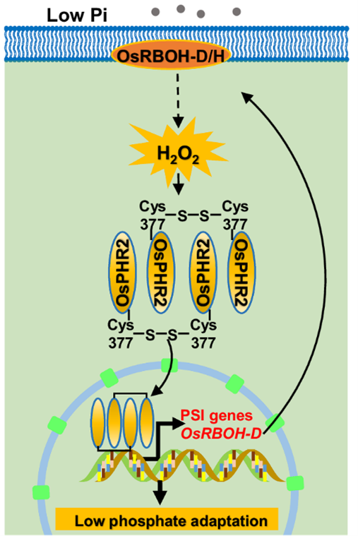 Phosphorus (P) is one of the indispensable macronutrients that fuel plant growth and development. However, most soil P is locked in insoluble complexes with heavy metals, making it largely inaccessible to plants. In rice, the MYB transcription factor PHOSPHATE STARVATION RESPONSE 2 (OsPHR2) acts as a central regulator of phosphate starvation-induced (PSI) genes to promote P uptake. Its activity is fine-tuned through intricate post-translational modifications and protein-protein interactions involving SPX proteins, inositol pyrophosphates (PP-InsPs), and SUMOylation. Intriguingly, Meng and colleagues uncovered a new messenger in phosphate starvation signaling: hydrogen peroxide (H₂O₂). Phosphate deficiency triggers H₂O₂ production, which in turn activates PSI gene expression. Loss of RBOH-D/H, enzymes responsible for H₂O₂ generation, impairs H₂O₂ accumulation, phosphate uptake, and plant biomass. Through BIAM labeling and site-directed mutagenesis, the team identified Cys377 of OsPHR2 as the key oxidation site mediating its oligomerization, nuclear translocation and DNA-binding activity. Moreover, OsPHR2 directly activates OsRBOH-D, establishing a positive feedback loop that amplifies phosphate starvation responses. This discovery not only redefines H₂O₂ as a pivotal messenger linking redox signaling to nutrient regulation but also opens new avenues for engineering crops with enhanced phosphate-use efficiency in the face of global resource constraints. (Summary by Ching Chan @ntnuchanlab) Nature Comms. 10.1038/s41467-025-63841-0
A plant virus uses plant metacaspase for its own benefit
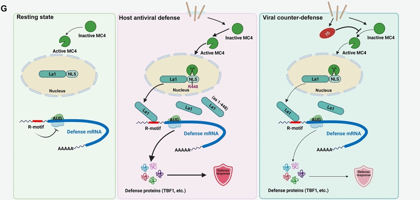 A recent study by Pi et al. decoded previously unknown mechanism through which a virus manipulates the plant defense pathway for successful infection. Focusing on the interaction between barley and the devastating barley stripe mosaic virus (BSMV), researchers identified a critical regulatory hub centered on the cleavage of a specific RNA binding protein. In an uninfected plant, a plant metacaspase, MC4, which can cleave a nucleus-localized protein La1, remains in an inactive form. During virus infection, MC4 is activated and translocates to the nucleus where it cleaves La1. The cleaved La1 now translocates to the cytoplasm where it interacts with the purine-rich R-motif found on some defense RNAs, and this interaction leads to cap-independent translation of host defense mRNAs and antiviral immunity. This specialized translational pathway is crucial because it allows the plant to rapidly synthesize essential defense proteins, bypassing the standard cap-dependent machinery that viruses often manipulate for their own replication. However, BSMV employs a counter defense mechanism in which the γb protein of the BSMV bind to MC4 and keeps it in inactivated state. Thus the cleavage of La1 is hampered and the cap-independent translation of host defense mRNA also hampered. These insights open new avenues for engineering durable viral resistance in crop species. (Summary by Ved Prakash @vedjiwan) Science Advances 10.1126/sciadv.adv0819
Illuminating plant immunity: A live sensor to watch salicylic acid in action
Salicylic acid (SA) is best known as a central hormone orchestrating plant immune response, including the hypersensitive reaction and systemic acquired resistance. Beyond defense, SA also influences plant growth and development, highlighting a delicate role in balancing the trade-off between immunity and productivity. To fully unravel how plants control these processes, tools are needed to track SA dynamics in real time and with cellular precision. This is a challenge that traditional methods, often destructive or indirect, have struggled to meet. To address this gap, Tang and colleagues developed a FRET-based sensor named SalicS1, designed specifically to visualize SA in living tissues. The sensor is built from a truncated Arabidopsis NPR1 protein linked to NIMIN1, which interact in vivo. SA binding to NPR1 disrupts this interaction, thereby decreasing the FRET emission ratio (negative ratio change sensor). By adding a nuclear localization signal, the improved version (nlsSalicS1) enabled stable in planta detection of SA across various organs, including roots, cotyledons, and mature leaves. Importantly, the sensor responsiveness can be validated through genetic manipulation: plants carrying NahG (which degrades SA) or mutations in EDS5 and PBS3 (which block SA synthesis) displayed notably lower FRET signals. Beyond its precision, nlsSalicS1 successfully detected SA responses triggered by diverse biotic stresses, from bacteria (Pseudomonas syringae), fungus (Blumeria graminis), and aphid (Brevicoryne brassicae). This powerful tool paves the way for live imaging of hormone dynamics and holds great promise for future applications in crop plants, bringing us closer to visualizing how plants fine-tune the balance between growth and defense in real time. (Summary by Ching Chan @ntnuchanlab) Science 10.1126/science.adw7650
The sweet connection between plants, fungi, and soil life
![]() Through their roots, plants live in close association with many soil microorganisms, including fungi and bacteria that help them grow. Beneficial fungi help plants cope with stress, and in return, the plants provide them with nutrients. A key part of this exchange is the movement of sucrose, a sugar produced by photosynthesis that fuels both plants and microbes. In this study, Fang and colleagues identified a new sucrose transporter called GspSUT1 in the fungus Gongronella butleri w5, a fungus that promotes plant growth and increases plant nutrient content such as nitrogen. This protein allows the fungus to take sucrose directly from plant roots and convert it into simple sugars such as glucose and fructose. When scientists silenced the GspSUT1 gene, they found more sucrose in the roots because the fungus was no longer absorbing it. Moreover, the growth-promoting effect of the fungus and the nitrogen content in the plant were reduced, disrupting the symbiotic interaction between the plant and the microorganism. The researchers also discovered that the glucose and fructose released into the soil feed nitrogen-fixing bacteria, microbes that convert atmospheric nitrogen into forms plants can use. As a result, plants have more nitrogen content, grow faster and healthier. Overall, GspSUT1 reveals a sophisticated cooperation between plants, fungi, and bacteria, showing how carbon and nitrogen cycles are tightly linked in healthy soils. Summary by Carlos González Sanz (@carlosgonzsanz). Current Biology (10.1016/j.cub.2025.08.043)
Through their roots, plants live in close association with many soil microorganisms, including fungi and bacteria that help them grow. Beneficial fungi help plants cope with stress, and in return, the plants provide them with nutrients. A key part of this exchange is the movement of sucrose, a sugar produced by photosynthesis that fuels both plants and microbes. In this study, Fang and colleagues identified a new sucrose transporter called GspSUT1 in the fungus Gongronella butleri w5, a fungus that promotes plant growth and increases plant nutrient content such as nitrogen. This protein allows the fungus to take sucrose directly from plant roots and convert it into simple sugars such as glucose and fructose. When scientists silenced the GspSUT1 gene, they found more sucrose in the roots because the fungus was no longer absorbing it. Moreover, the growth-promoting effect of the fungus and the nitrogen content in the plant were reduced, disrupting the symbiotic interaction between the plant and the microorganism. The researchers also discovered that the glucose and fructose released into the soil feed nitrogen-fixing bacteria, microbes that convert atmospheric nitrogen into forms plants can use. As a result, plants have more nitrogen content, grow faster and healthier. Overall, GspSUT1 reveals a sophisticated cooperation between plants, fungi, and bacteria, showing how carbon and nitrogen cycles are tightly linked in healthy soils. Summary by Carlos González Sanz (@carlosgonzsanz). Current Biology (10.1016/j.cub.2025.08.043)
Late ROS burst as a signature of exotoxin-triggered immunity in plants
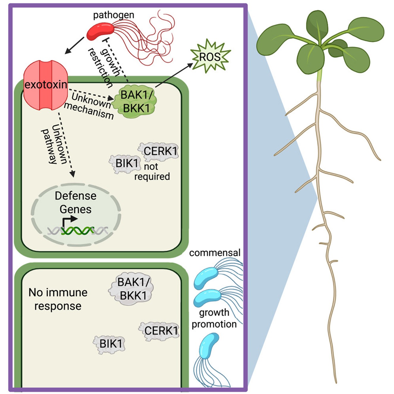 Plants depend on innate immune systems to recognize microbial invaders or damages that signal danger. This discrimination must be extremely precise: they must welcome beneficial microbes that support nutrient uptake and growth while defending against pathogens that threaten survival. A long-standing question in plant-microbe interactions is how plants distinguish between closely related beneficial and pathogenic bacteria that share similar molecular signatures. Thoms and colleagues tackled this challenge using two phylogenetically related Pseudomonas fluorescens strains associated with plant roots. The beneficial strain WCS365 promotes plant growth, whereas the pathogenic strain N2C3 causes disease. Furthermore, the pathogenic strain but not the beneficial strain produces small secreted lipopeptide exotoxins. Through the use of fluorescent reporter lines and exotoxin-deficient bacterial mutants, the researchers identified the N2C3-produced exotoxin as the key determinant of immune activation and pathogen suppression. Surprisingly, the immune response triggered by this exotoxin operates independently of conventional hormone signaling pathways. Instead, it induces a delayed oxidative burst, distinct from the rapid ROS production elicited by the canonical flg22-triggered pattern-triggered immunity. The coreceptors BAK1 and BKK1 are critical for this toxin-induced ROS production as for flg22. This study reveals that plants can distinguish between nearly identical microbial relatives by detecting the presence of specific exotoxins, highlighting a sophisticated immune surveillance system. (Summary by Ching Chan @ntnuchanlab) Cell Reports 10.1016/j.celrep.2025.116457
Repeated evolution of flowers specialized for buzz pollination
 If you’ve ever looked closely at a tomato flower, you might have noticed that its anthers cluster together in the center of the flower, making the pollen inaccessible to most insects. Tomato flowers are an example of a buzz pollinated flower, in which the pollen is released by the physical shaking by a pollinator (usually but not always a bee), and the pollen falls through a small pore where it is collected by the bee (check out this YouTube video to see it in action). Tomato flowers are a type of poricidal flowers, not all of which are buzz pollinated, but which share the characteristic of having a small pore though which pollen is released. Poricidal flowers are found throughout the angiosperms, so Russel et al. decided to look at their phylogenetic distribution and evolutionary origins. They found that about 10% of angiosperm species in at least 639 genera have the poricidal morphology, and that this morphology has at least 205 distinct evolutionary origins. The trait may be beneficial in some environments (e.g., arid and with low wind) but it has the disadvantage of being more dependent on specific pollinators. Based on their phylogenetic data, the authors conclude that the transition to poricidal morphology is rare, but the probability of losing it is high. As yet the developmental programs underpinning this fascinating evolutionary convergence remain to be identified. And, watching a bee quiver and shake to capture some pollen is a great way to get people interested in plant science! (Summary by Mary Williams @PlantTeaching.bsky.social) Evolution 10.1093/evolut/qpaf220.
A vision for plant science education
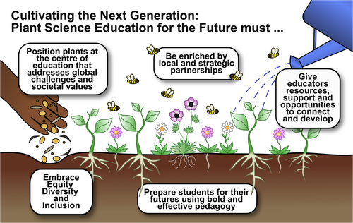 The urgent need to support and sustain plant research is indisputable, but sometimes plant education, the foundation upon which research rests, doesn’t get the same support. In January 2025, a group of dedicated educators (mostly higher education and outreach) spent two days together to strategize a vision for a secure, sustainable, and effective educational landscape. The result, titled a Manifesto for Plant Science Education, identified five themes: (i) plants must be at the center of an education that addresses global challenges and societal values; (ii) plant science education must prepare students for their futures using bold and effective pedagogies; (iii) equity, diversity and inclusion must be robustly embedded in educational practices; (iv) local and strategic partnerships (with industry and beyond) are required to strengthen academic education; and (v) plant science educators need resources and opportunities to develop and connect. Specific actions in support of the five themes are provided. The authors conclude with a call to action, “Just as investment in plant science research is deemed essential and urgent, so too is a concerted and sustained commitment to plant science education at all levels. We urge educators, institutions, policymakers and industry partners to actively engage with the recommendations made here.” (Summary by Mary Williams @PlantTeaching.bsky.social) Plants People Planet 10.1002/ppp3.70115

