Metabolites through the looking glass with CEST MRI
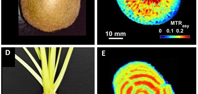
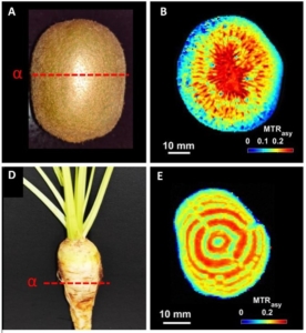 Non-invasive imaging technologies like computed tomography and magnetic resonance imaging (MRI) have revolutionized medicine by improving diagnostics and guiding treatment. Due to its versatility, MRI also holds potential for plant sciences, where it can be used to visualize and quantify metabolites within organs, tissues, and cells. However, challenges specific to plant tissues have hindered its implementation. Mayer et al. addressed these challenges by integrating chemical exchange saturation transfer (CEST), an MRI contrast technique that enhances signal detection and reduces susceptibility to magnetic field disturbances in plant tissues. CEST MRI enables high-resolution visualization of metabolites like amino acids and sugars. The researchers tested their method on various plant systems, from model crops like potato tubers to more complex tissues such as maize kernels. Compared to standard MRI, CEST MRI offers a higher signal-to-noise ratio, better spatial resolution, and faster detection times. Additionally, its ability to image live and complex plant tissues, such as barley grains on the spike, make it a powerful tool for the in vivo detection of metabolites in economically important sink organs including tubers, roots, and grains. Unlike most metabolomics techniques, which measure specific metabolites destructively, CEST MRI captures broad metabolite classes non-invasively, in live tissues, and with high resolution. This remarkable advancement in the field of plant metabolism has exciting potential for plant phenotyping platforms, breeding programs, and biofortification strategies by selecting traits linked to optimal metabolite concentrations. (Summary by Thomas Depaepe @thdpaepe.bsky.social @thdpaepe) Science Advances 10.1126/sciadv.adq4424


